. 2024 Oct 30;10(1):34.
doi: 10.1186/s41205-024-00241-y.

An anthropomorphic phantom for atrial transseptal puncture simulation training

Affiliations

An anthropomorphic phantom for atrial transseptal puncture simulation training

Aya Mutaz Zeidan et al. 3D Print Med. .

Abstract

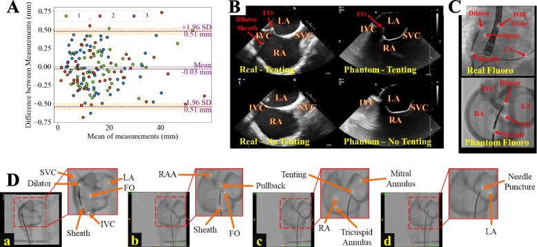
Background: Transseptal puncture (TSP) is a critical prerequisite for left-sided cardiac interventions, such as atrial fibrillation (AF) ablation and left atrial appendage closure. Despite its routine nature, TSP can be technically demanding and carries a risk of complications. This study presents a novel, patient-specific, anthropomorphic phantom for TSP simulation training that can be used with X-ray fluoroscopy and ultrasound imaging.

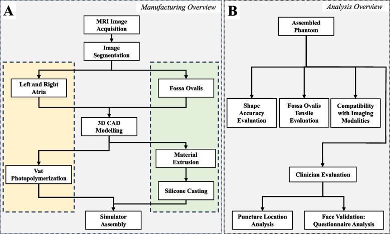
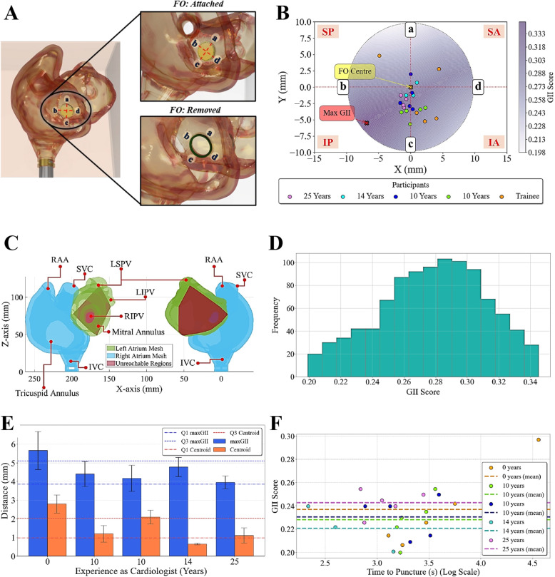
Methods: The TSP phantom was developed using additive manufacturing techniques and features a replaceable fossa ovalis (FO) component to allow for multiple punctures without replacing the entire model. Four cardiologists and one cardiology trainee performed TSP on the simulator, and their performance was assessed using four metrics: global isotropy index, distance from the centroid, time taken to perform TSP, and a set of 5-point Likert scale questions to evaluate the clinicians' perception of the phantom's realism and utility.

Results: The results demonstrate the simulator's potential as a training tool for interventional cardiology, providing a realistic and controllable environment for clinicians to refine their TSP skills. Experienced cardiologists tended to cluster their puncture points closer to regions of the FO associated with higher global isotropy index scores, indicating a relationship between experience and optimal puncture localization. The questionnaire analysis revealed that participants generally agreed on the phantom's realistic anatomical representation and ability to accurately visualize the TSP site under fluoroscopic guidance.

Conclusions: The TSP simulator can be incorporated into training programs, offering trainees the opportunity to improve tool handling, spatial coordination, and manual dexterity prior to performing the procedure on patients. Further studies with larger sample sizes and longitudinal assessments are needed to establish the simulator's impact on TSP performance and patient outcomes.

Keywords: 3D printing; Cardiology; Patient-specific; Simulation; Training; Transseptal puncture.


Conflict of interest statement

The authors declare no competing interests.

Figures

Fig. 1
Fig. 1
Phantom manufacturing and analysis overview of a patient-specific TSP simulator. Yellow: biatrial structure. Green: Replaceable FO. Abbreviations: TSP (transseptal puncture); MRI (magnetic resonance imaging); CAD (computer-aided design); FO (fossa ovalis)
Fig. 2
Fig. 2
Mapping the segmentation of the LA (green) and RA (blue) based on a cardiac MRI scan of an AF patient. A Sagittal view. B Transverse view. C 3D visualization, including: (a) RAA, (b) SVC, (c) RSPV, (d) RIPV, (e) LSPV, and (f) LIPV. D, E and F, G visualize CAD renderings and images of the fabricated phantom in two oblique views. Abbreviations: RA (right atrium); LA (left atrium); MRI (magnetic resonance imaging); AF (atrial fibrillation); RAA (right atrial appendage); SVC (superior vena cava), IVC (inferior vena cava); RSPV (right superior pulmonary vein); RIPV (right inferior pulmonary vein); LSPV (left superior pulmonary vein); LIPV (left inferior pulmonary vein); CAD (computer-aided design); FO (fossa ovalis); PVC (polyvinyl chloride)
Fig. 3
Fig. 3
FO modeling and characterization. A Silicone mold of the FO based on patient-specific MRI segmentation, showing four key measurements: (a) maximum FO thickness, (b) FO diameter, (c) diameter of the interchangeable FO clamp, and (d) diameter of the molded silicone FO. B The resulting silicone FO insert after casting. C Comparison of mechanical properties of various silicone mixtures considered for FO development, including the mean Young’s modulus, mean tensile stress at break, and mean tensile extension at break. Abbreviations: MRI (magnetic resonance imaging); FO (fossa ovalis); EF (EcoFlex); SLK (slacker): S (superior); I (inferior); A (anterior); P (posterior)
Fig. 4
Fig. 4
Simulator assembly. A Watertight assembly: The phantom was enclosed in a transparent box that facilitates X-ray fluoroscopy and ultrasound imaging by filling the box with water. (a) A PVC tube was attached from the phantom to a socket that was (b) secured to the box with (c) screws and O-rings placed on either side of the box and the (d) femoral vein socket and corresponding PVC tube, which was closed off with a (e) tear-resistant silicone adapter. B Phantom Assembly: (f) socket adapter and (g) PVC tube secured to the (h) phantom fitted with a permanent (i) FO ring and (j) replaceable FO insert. C FO attachment. D Complete assembly. Abbreviations: PVC (polyvinyl chloride); FV (femoral vein); FO (fossa ovalis); BRK (Brockenbrough); RA (right atrium); LA (left atrium); IVC (inferior vena cava)
Fig. 5
Fig. 5
Experimental setup for the pilot study. A Adult catheterization laboratory, where the tools were tracked using (a) an Aurora EM-Field Generator and guided by (b) real-time X-ray fluoroscopy. B A cardiologist performing TSP using a (c) BRK-1 needle and (d) dilator-sheath. C Phantom fitted with (e) a reference sensor fastened to the cardiac structure above the (g) FO insert and a (f) sensor attached to the dilator tip. The phantom sits on a (h) mesh as the chest cavity. Abbreviations: EM (electromagnetic); TSP (transseptal puncture); BRK (Brockenbrough); FO (fossa ovalis)
Fig. 6
Fig. 6
Imaging compatibility and shape accuracy evaluation of the patient-specific phantom. A Bland-Altman plot assessing the agreement between the dimensions of the STL model and the fabricated phantom. B Ultrasound imaging of the phantom, demonstrating its visualization of key anatomical structures, such as the FO, is essential for accurate TSP performance. Real ultrasound scans were obtained in the TOE bicaval view. C Real and phantom X-ray fluoroscopy images. D Real-time fluoroscopic guidance depicting critical steps in the TSP procedure, including (a, b) positioning of the TSP kit in the SVC in the RAO 30 and LAO 60 projections, respectively, (c) tenting of the fossa ovalis (FO), and (d) needle puncture. Abbreviations: STL (stereolithography); TSP (transseptal puncture); FO (fossa ovalis); SVC (superior vena cava); IVC (inferior vena cava); RAO (right anterior oblique); LAO (left anterior oblique); RA (right atrium); LA (left atrium); RAA (right atrial appendage); CS (coronary sinus)
Fig. 7
Fig. 7
Puncture positioning. A Orientation of the FO and its division: (a), (b), (c), and (d) correspond to superior, posterior, inferior, and anterior orientations of the FO based on the schematic divisions (SP, IP, SA, and IA) defined in [9]. B GII scores and participant puncture locations on the FO mesh. C Regions on the LA unreachable by the catheter, given the catheter pivots about the patient-specific optimal puncture location. D Histogram of the frequency of GIIs. D GII scores against the time taken to perform TSP. E Mean Euclidean distance of participants’ puncture locations from the maximum GII score and FO centroid coordinates. F GII scores against the logarithmic scale of time. Abbreviations: FO (fossa ovalis); SP (superior-posterior); IP (inferior-posterior); SA (superior-anterior); IA (inferior-anterior); GII (global isotropy index)

References

    1. Vinter N, Huang Q, Fenger-Grøn M, Frost L, Benjamin EJ, Trinquart L. Trends in excess mortality associated with atrial fibrillation over 45 years (Framingham Heart Study): community based cohort study. BMJ. 2020;370:1–8. - PMC - PubMed
    1. Krijthe BP, Kunst A, Benjamin EJ, Lip GYH, Franco OH, Hofman A, et al. Projections on the number of individuals with atrial fibrillation in the European Union, from 2000 to 2060. Eur Heart J. 2013;34(35):2746–51. - PMC - PubMed
    1. De Ponti R, Marazzi R, Ghiringhelli S, Salerno-Uriarte JA, Calkins H, Cheng A. Superiority of simulator-based training compared with conventional training methodologies in the performance of transseptal catheterization. J Am Coll Cardiol. 2011;58(4):359–63. - PubMed
    1. Alkhouli M, Rihal CS, Holmes DR. Transseptal techniques for emerging structural heart interventions. JACC Cardiovasc Interv. 2016;9(24):2465–80. - PubMed
    1. Naik N. How to perform transeptal puncture. Indian Heart J. 2015;67(1):70–6. - PMC - PubMed

LinkOut - more resources