. 2024 Oct 29;7(1):1410.
doi: 10.1038/s42003-024-07100-w.

Two ubiquitous aldo-keto reductases in the genus Papaver support a patchwork model for morphine pathway evolution

Affiliations

Two ubiquitous aldo-keto reductases in the genus Papaver support a patchwork model for morphine pathway evolution

Samuel C Carr et al. Commun Biol. .

Abstract

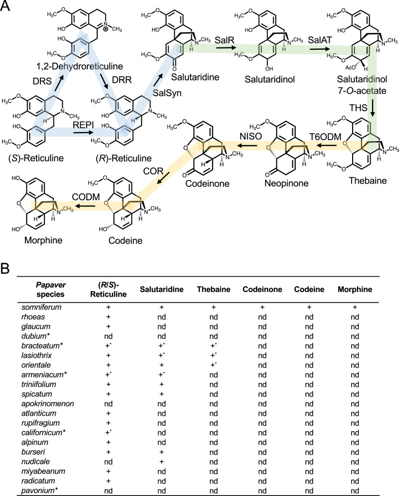
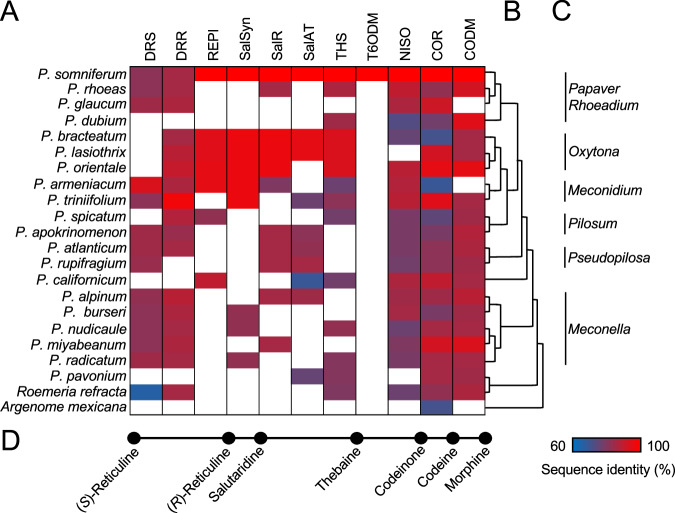
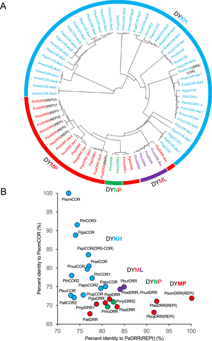
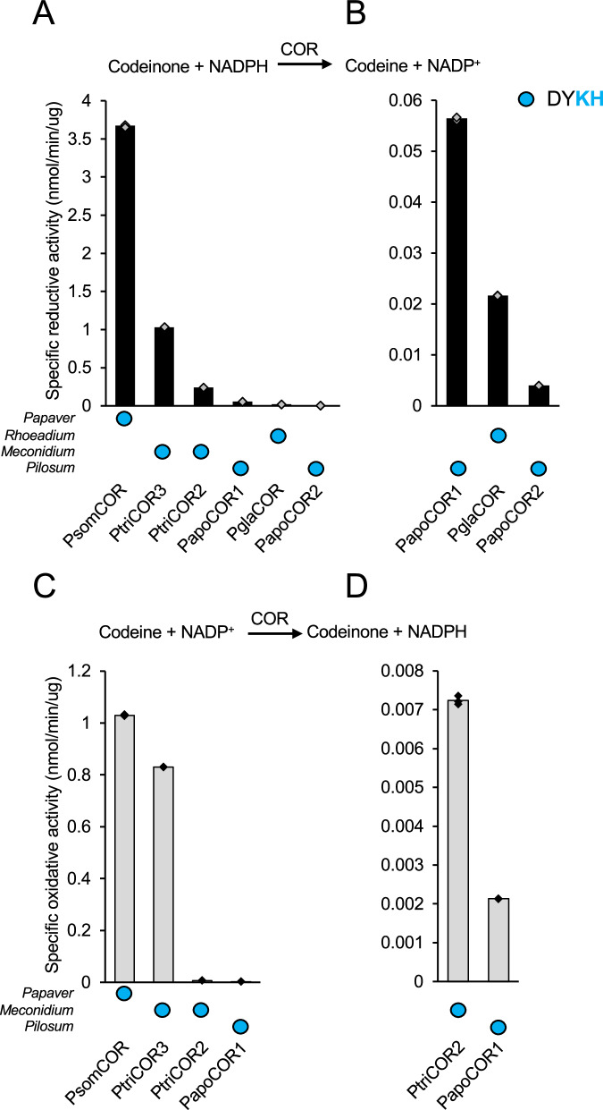
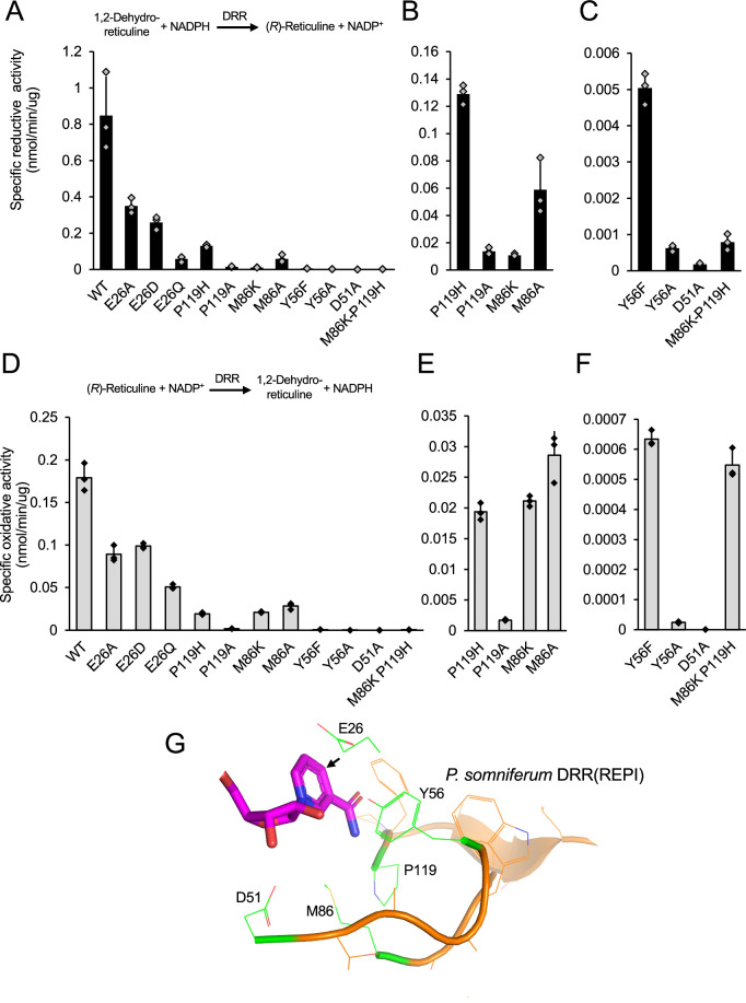
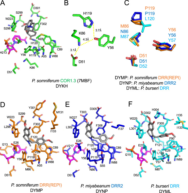
The evolution of morphinan alkaloid biosynthesis in plants of the genus Papaver includes permutation of several processes including gene duplication, fusion, neofunctionalization, and deletion resulting in the present chemotaxonomy. A critical gene fusion event resulting in the key bifunctional enzyme reticuline epimerase (REPI), which catalyzes the stereochemical inversion of (S)-reticuline, was suggested to precede neofunctionalization of downstream enzymes leading to morphine biosynthesis in opium poppy (Papaver somniferum). The ancestrally related aldo-keto reductases 1,2-dehydroreticuline reductase (DRR), which occurs in some species as a component of REPI, and codeinone reductase (COR) catalyze the second and penultimate steps, respectively, in the pathway converting (S)-reticuline to morphine. Orthologs for each enzyme isolated from the transcriptomes of 12 Papaver species were shown to catalyze their respective reactions in species that capture states of the metabolic pathway prior to key evolutionary events, including the gene fusion event leading to REPI, thus suggesting a patchwork model for pathway evolution. Analysis of the structure and substrate preferences of DRR orthologs in comparison with COR orthologs revealed structure-function relationships underpinning the functional latency of DRR and COR orthologs in the genus Papaver, thus providing insights into the molecular events leading to the evolution of the pathway.


Conflict of interest statement

The authors declare no competing interests.

Figures

Fig. 1
Fig. 1. Alkaloid profiling of various Papaver species.
A Morphinan alkaloid biosynthetic pathway in opium poppy. Steps from (S)-reticuline to salutaridine are highlighted in blue, the pathway from salutaridine to thebaine is highlighted in green, and the conversion of thebaine to morphine is highlighted in yellow. B Alkaloids in the pathway from (S)-reticuline to morphine detected in various Papaver species. ‘+’ indicates that the corresponding alkaloid was identified and the abbreviation ‘nd’ indicates that no alkaloid was detected. ‘+*’ indicate detected alkaloids reported in other published work,.
Fig. 2
Fig. 2. Transcript profiling of various Papaver species.
A Occurrence of transcripts encoding proteins exhibiting greater than 60% amino acid sequence identity relative to enzymes involved in the conversion of (S)-reticuline to morphine in opium poppy. Nucleotide sequencing and contig assembly statistics are provided for each transcriptome in Table S8. Transcripts showing relatively higher predicted amino acid sequence identity with a different enzyme were omitted. Numerical percent identities and indication of partial and truncated transcript with the corresponding percent coverage are provided in Fig S2. Relative amino acid sequence identity with the corresponding opium poppy biosynthetic enzyme is shown as a gradient from red (100%) to blue (60%). White indicates that no transcript was annotated. Transcriptomes from previously published work were used for Papaver rhoeas,, P. dubium P. bracteatum P. orientale P. armeniacum P. californicum and P. pavonium, R. refracta, and A. mexicana. B Consensus phylogenetic relationship among Papaver species based on previous published work,. C Selected section denominations of Papaver species. D Simplified diagram of the morphine biosynthetic pathway showing key metabolites from Fig. 1B and their corresponding position in panel A.
Fig. 3
Fig. 3. Phylogenetic and sequence similarity analysis of AKR homologs from various Papaver species.
A Phylogenetic tree showing the relationships among full-length AKR transcripts found in transcriptomes from various Papaver species. Annotations of ‘COR-like’ or ‘DRR-like’ was based on the catalytic tetrad found in each predicted polypeptide. COR-like proteins containing the DYKH catalytic tetrad are shown in cyan, whereas DRR-like proteins are indicated in red (DYMP), purple (DYML), or green (DYNP). The function of transcripts lacking a ‘like’ suffix have been empirically confirmed. AKRs occurring as a fusion with a cytochrome P450 (i.e., a REPI homolog) are shown in black. The neighbor-joining tree was constructed using the Geneious tree-building Tamura-Nei model with a cost matrix of 65% similarity (5.0/−4.0), a gap open-penalty of 12, and a gap extension-penalty of 3 (nodes: 136; tips: 69). B Sequence similarity analysis of selected AKRs found in transcriptome from various Papaver species. Predicted proteins were plotted based on amino acid sequence identity compared with COR and DRR (from REPI), calculated using Clustal Omega. COR-like proteins containing the DYKH catalytic tetrad are shown in cyan, whereas DRR-like proteins are indicated in red (DYMP), purple (DYML), or green (DYNP).
Fig. 4
Fig. 4. Functional characterization of DRR homologs from various Papaver species.
Specific activities of select DRR homologs. Corresponding enzyme Papaver sections and catalytic tetrad motifs are shown underneath the X-axis and the reaction coordinate above the plots. A,B Black bars indicate specific activity in ‘forward’ reductive enzyme assays measuring the formation of (R)-reticuline from 20 μM 1,2-dehydroreticuline and 500 μM NADPH. Reactions were carried out in 100 mM Bis-tris propane buffer, pH 7.0. A Data for all characterized DRRs. B Data for a subset of DRRs with relatively low reductive activity. C,D Gray bars indicate specific activity in ‘reverse’ oxidative enzyme assays measuring the formation of 1,2-dehydroreticuline from 20 μM (R)-reticuline and 500 μM NADP+. Reactions were carried out in 100 mM Bis-tris propane buffer, pH 8.8. C Data for all characterized DRRs. D Data for a subset of DRRs with relatively low oxidative activity. The protein concentrations and reaction times required to achieve less than 10% substrate conversion for each enzyme are provided in Table S10. Bars represent the mean ± standard deviation of three independent replicates.
Fig. 5
Fig. 5. Functional characterization of COR homologs from various Papaver species.
Specific activities of select COR homologs. Corresponding enzyme Papaver sections and catalytic tetrad motifs are shown underneath the X-axis and the reaction coordinate above the plots. A,B Black bars indicate specific activity in ‘forward’ reductive enzyme assays measuring the formation of codeinone from 50 μM codeine and 1 mM NADPH. Reactions were carried out in 100 mM Bis-tris propane buffer, pH 8.0. A Data for all characterized CORs. B Data for a subset of CORs with relatively low reductive activity. C,D Gray bars indicate specific activity in ‘reverse’ oxidative enzyme assays measuring the formation of 1,2-dehydroreticuline from 50 μM codeine and 1 mM NADP+. Reactions were carried out in 100 mM Bis-tris propane buffer, pH 8.8. C Data for all characterized CORs. D Data for a subset of CORs with relatively low oxidative activity. The protein concentrations and reaction times required to achieve less than 10% substrate conversion for each enzyme are provided in Table S10. Bars represent the mean ± standard deviation of three independent replicates.
Fig. 6
Fig. 6. Mutagenesis of the DRR component of REPI from opium poppy reveals a novel catalytic tetrad.
AC Black bars indicate specific activity of DRR mutants in ‘forward’ reductive enzyme assays measuring the formation of (R)-reticuline from 20 μM 1,2-dehydroreticuline and 500 μM NADPH. Reactions were carried out in 100 mM Bis-tris propane buffer, pH 7.0. A Data for all characterized DRRs. B Data for a subset of DRRs with relatively moderate reductive activity. C Data for a subset of DRRs with relatively low reductive activity. DF Gray bars indicate specific activity of DRR mutants in ‘reverse’ oxidative enzyme assays measuring the formation of 1,2-dehydroreticuline from 20 μM (R)-reticuline and 500 μM NADP+. Reactions were carried out in 100 mM Bis-tris propane buffer, pH 8.8. D Data for all characterized DRRs. E Data for a subset of DRRs with relatively moderate oxidative activity. F Data for a subset of DRRs with relatively low oxidative activity. The protein concentrations and reaction times required to achieve less than 10% substrate conversion for each enzyme are provided in Table S11. Bars represent the mean ± standard deviation of three independent replicates. Reaction coordinates are shown above the plots. G Depiction of the active site residues selected for mutagenesis from the homology model of P. somniferum DRR from REPI. Mutagenesis candidate residues are shown in green, NADPH in magenta, and the pro R face of NADPH is designated with an arrow.
Fig. 7
Fig. 7. Characterization of the binding pocket in AKRs from various Papaver species.
A Crystal structure of COR (7MBF) from opium poppy (P. somniferum) shown in green. B Arrangement of the catalytic tetrad in COR from P. somniferum. C Superposition of the respective motifs of substitutions to the canonical AKR catalytic tetrad in P. somniferum DRR(REPI) (orange), P. miyabeanum DRR2 (blue), and P. burseri DRR (cyan). D the DRR component of REPI from P. somniferum shown in orange, (E) DRR2 from P. miyabeanum shown in blue, and F DRR from P. burseri shown in cyan. In all cases, a docked molecule of NADPH is shown in magenta and docked ligand molecules (i.e., codeinone for COR and 1,2-dehydroreticuline for DRR) are shown in gray. Carbon atoms are represented by the base color of each enzyme, with red corresponding to oxygen, blue to nitrogen, and yellow to sulfur. Hydrogen bonding and electrostatic interactions are shown in yellow (predicted) and red (disrupted) dashed lines. Red arrows depict predicted movements of sidechains and substrates based on binding pocket residue substitutions. PyMOL was used to generate images of the models.
Fig. 8
Fig. 8. Functional characterization of DRR, DRS, REPI, and SalSyn homologs from various Papaver species in engineered yeast strains.
A DRS, (B,C) SalSyn, (D) DRR, (E) REPI and unfused DRS-DRR pairs were transiently expressed in one of two yeast strains: (i) a BUP1- and CPR2-chromosomally integrated strain (used for DRS, SalSyn and DRR) and (ii) a BUP1-, CPR2- and SalSyn-chromosomally integrated strain (used for REPI, and unfused DRS and DRR pairs). Yeast strains were feed 50 μM of the indicated substrate (i.e., (S)-reticuline for DRS, (R)-reticuline for SalSyn, 1,2-dehydroreticuline for DRR, and (S)-reticuline for REPI, and unfused DRS and DRR pairs. Yeast cultures were incubated for the indicated time (i.e., 12, 24 or 48 h) and conversion products (i.e., 1,2-dehydroreticuline for DRS, salutaridine for SalSyn, (R)-reticuline for DRR, and salutaridine, (S)-reticuline and 1,2-dehydroreticuline for REPI, and unfused DRS and DRR pairs) were detected and quantified using a five-point standard curve. Error bars correspond to the mean ± standard deviation of three biological replicates (i.e., three independent colonies). The catalyzed reaction and sequences are shown above each plot.

References

    1. Dastmalchi, M., Park, M. R., Morris, J. S. & Facchini, P. Family portraits: the enzymes behind benzylisoquinoline alkaloid diversity. Phytochem. Rev.17, 249–277 (2018).
    1. World Health Organization. World health organization model list of essential medicines. Ment. Holist. Health.: Some Int. Perspect.21, 119–134 (2019).
    1. Board, I. N. C. Narcotic Drugs - Technical Report. (2019).
    1. Farrow, S. C., Hagel, J. M., Beaudoin, G. A. W., Burns, D. C. & Facchini, P. J. Stereochemical inversion of (S)-reticuline by a cytochrome P450 fusion in opium poppy. Nat. Chem. Biol.11, 728–732 (2015). - PubMed
    1. Winzer, T. et al. Morphinan biosynthesis in opium poppy requires a P450-oxidoreductase fusion protein. Science349, 309–312 (2015). - PubMed

LinkOut - more resources