

Identification of a disulfidptosis-related genes signature for diagnostic and immune infiltration characteristics in endometriosis
- PMID: 39472502
- PMCID: PMC11522465
- DOI: 10.1038/s41598-024-77539-8
Identification of a disulfidptosis-related genes signature for diagnostic and immune infiltration characteristics in endometriosis
Abstract
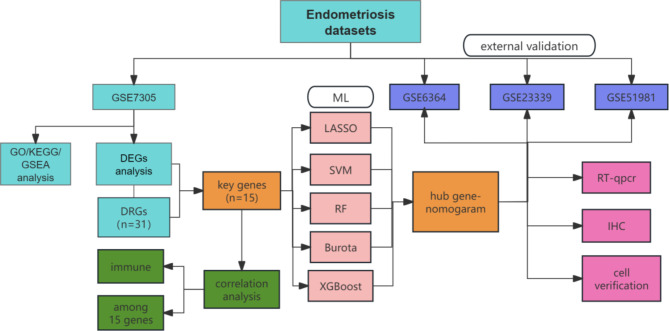
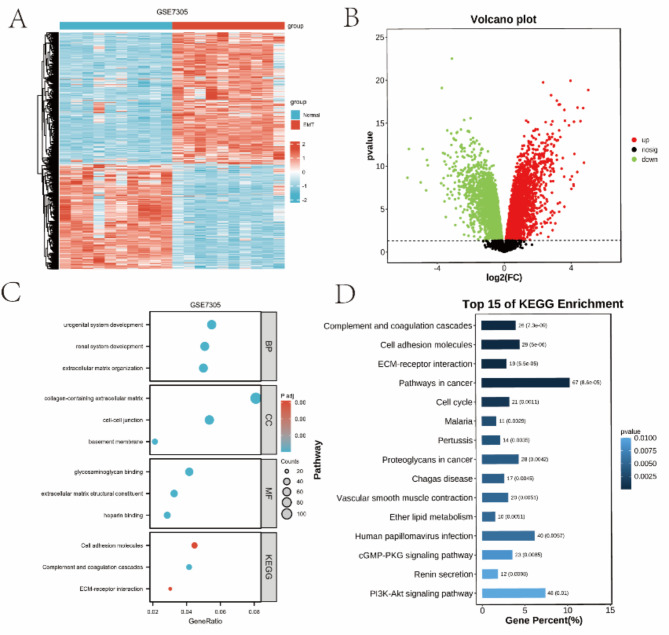
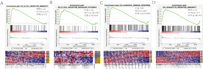
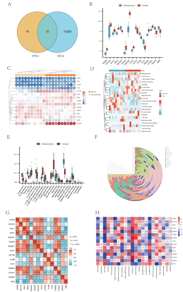
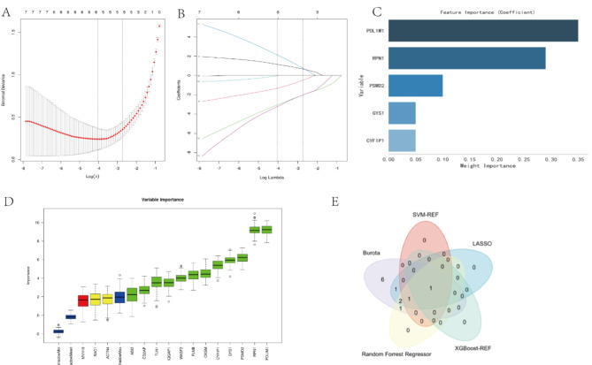
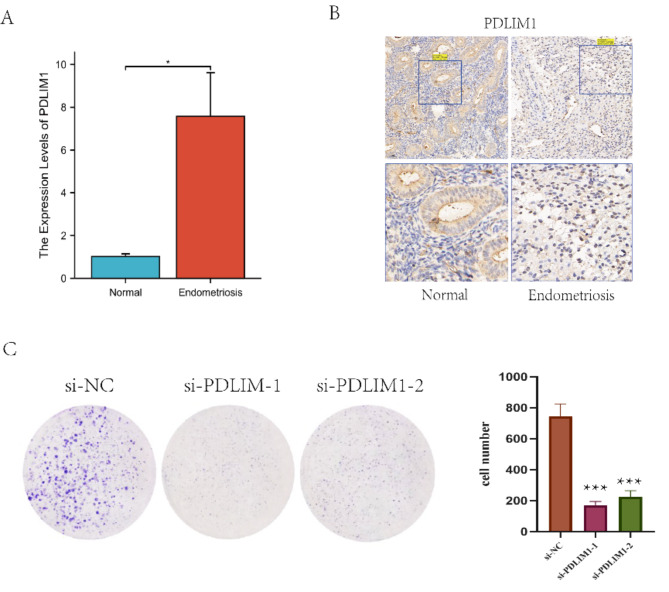
Endometriosis (EMs) is the prevalent gynecological disease with the typical features of intricate pathogenesis and immune-related factors. Currently, there is no effective therapeutic intervention for EMs. Disulfidptosis, the cell death pattern discovered recently, may show close relationships to immunity and EMs. In this study, bioinformatics analysis was used to investigate the role of disulfide breakdown related genes (DRGs) in EMs. The EMs gene expression matrix was subjected to differential analysis for identifying overlap between differentially expressed genes (DEGs) in EMs and genes associated with disulfide poisoning. Immunoinfiltration analysis was performed. In addition, the association of hub genes with immune cells was examined. Multiple machine learning methods were employed to identify hub genes, construction of predictive models, and validation using external datasets and clinical samples. Totally 15 overlapping genes were identified. Immune-correlation analysis showed that NK cells played a vital role, and these 15 genes were closely related to NK cells. PDLIM1 was further determined as the hub gene through machine learning techniques. Clinical samples and external datasets were adopted for validating the performance in diagnosis. According to the above findings, we built the predictive model, and calculated the AUCs obtained from three external validation datasets to demonstrate the model accuracy. RT-qPCR and IHC analyses were applied to confirm the results. Colony formation was used to verify the effect of PDLIM1 on the proliferation of primary EMs cells. A strong correlation between disulfidptosis and EMs was identified in this study, highlighting its close correlation with the immune microenvironment. Moreover, our results shed new lights on exploring biomarkers and potential therapeutic targets for EMs.
Keywords: Biomarkers; Disulfidptosis; Endometriosis; Immune.
© 2024. The Author(s).
Conflict of interest statement
The authors declare no competing interests.
Figures

Similar articles
-
Unraveling pathogenesis, biomarkers and potential therapeutic agents for endometriosis associated with disulfidptosis based on bioinformatics analysis, machine learning and experiment validation.J Biol Eng. 2024 Jul 26;18(1):42. doi: 10.1186/s13036-024-00437-0. J Biol Eng. 2024. PMID: 39061076 Free PMC article.
-
Identification of a disulfidptosis-related genes signature for diagnostic and immune infiltration characteristics in cervical cancer.PLoS One. 2025 Apr 30;20(4):e0322387. doi: 10.1371/journal.pone.0322387. eCollection 2025. PLoS One. 2025. PMID: 40305445 Free PMC article.
-
Shared diagnostic biomarkers and underlying mechanisms between endometriosis and recurrent implantation failure.Front Endocrinol (Lausanne). 2025 Feb 19;16:1490746. doi: 10.3389/fendo.2025.1490746. eCollection 2025. Front Endocrinol (Lausanne). 2025. PMID: 40046872 Free PMC article.
-
Integrated bioinformatic analysis of immune infiltration and disulfidptosis related gene subgroups in type A aortic dissection.Sci Rep. 2025 Apr 21;15(1):13719. doi: 10.1038/s41598-025-98149-y. Sci Rep. 2025. PMID: 40258895 Free PMC article.
-
Role of neutrophils in the immune microenvironment of endometriosis.Afr J Reprod Health. 2025 May 16;29(5s):13-19. doi: 10.29063/ajrh2025/v29i5s.2. Afr J Reprod Health. 2025. PMID: 40387046 Review.
References
-
- Taylor, H. S., Kotlyar, A. M. & Flores, V. A. Endometriosis is a chronic systemic disease: clinical challenges and novel innovations. Lancet397, 839–852 (2021). - PubMed
-
- Othman Eel, D. et al. Serum cytokines as biomarkers for nonsurgical prediction of endometriosis. Eur. J. Obstet. Gynecol. Reprod. Biol.137, 240–246 (2008). - PubMed
MeSH terms
Grants and funding
LinkOut - more resources
Full Text Sources
Medical
Research Materials
Miscellaneous
