. 2024 Oct 29;7(1):1408.
doi: 10.1038/s42003-024-07105-5.

Neuronal excitation-inhibition imbalance in the basolateral amygdala is involved in propofol-mediated enhancement of fear memory

Affiliations

Neuronal excitation-inhibition imbalance in the basolateral amygdala is involved in propofol-mediated enhancement of fear memory

Chen Chen et al. Commun Biol. .

Abstract

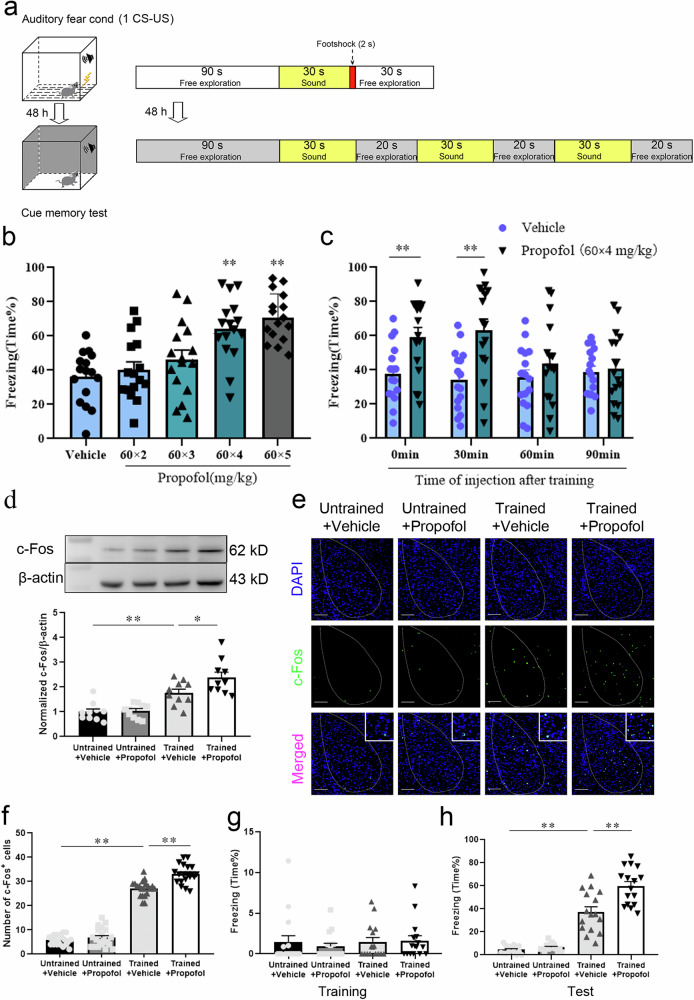
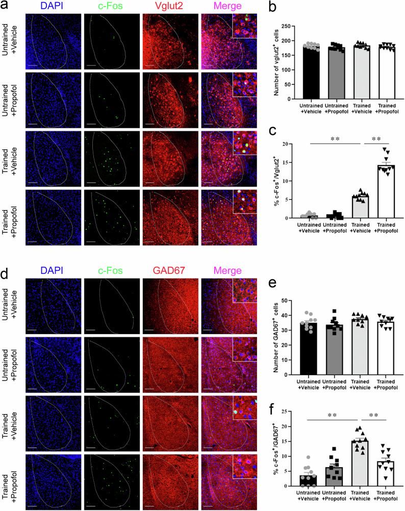
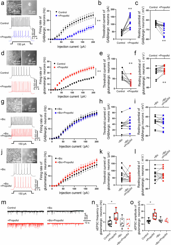
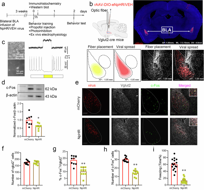
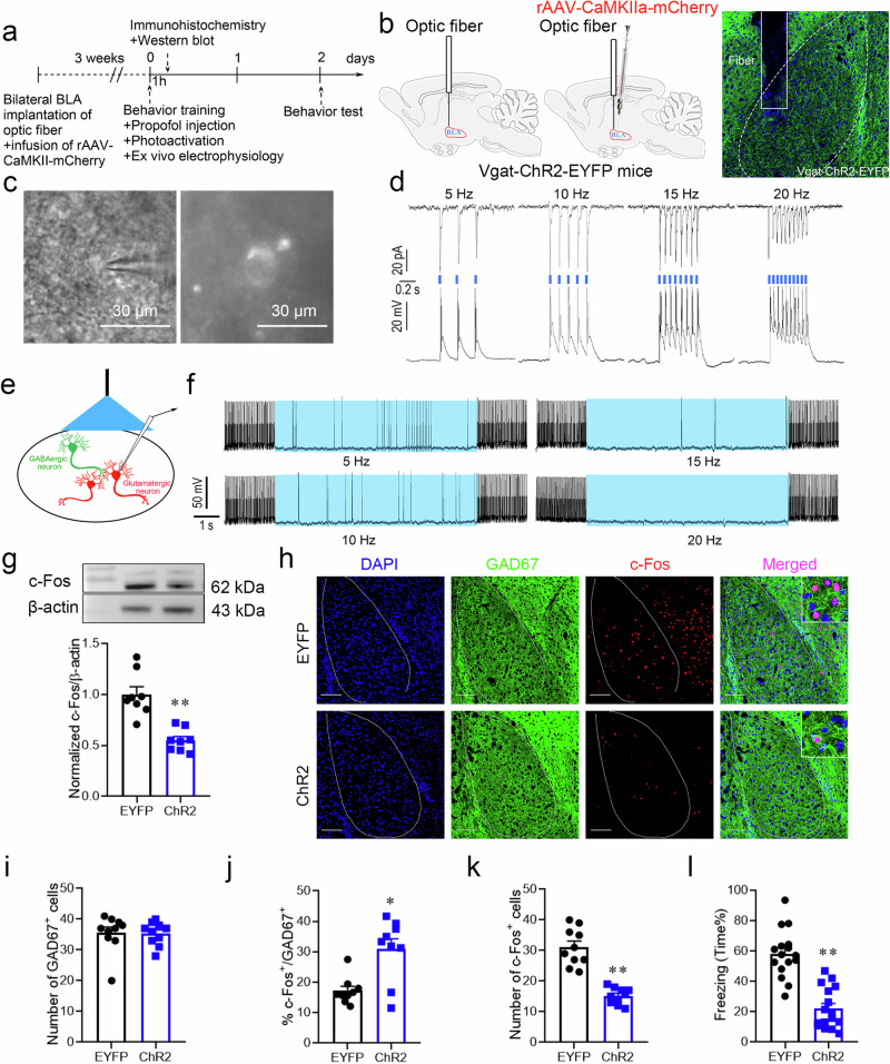
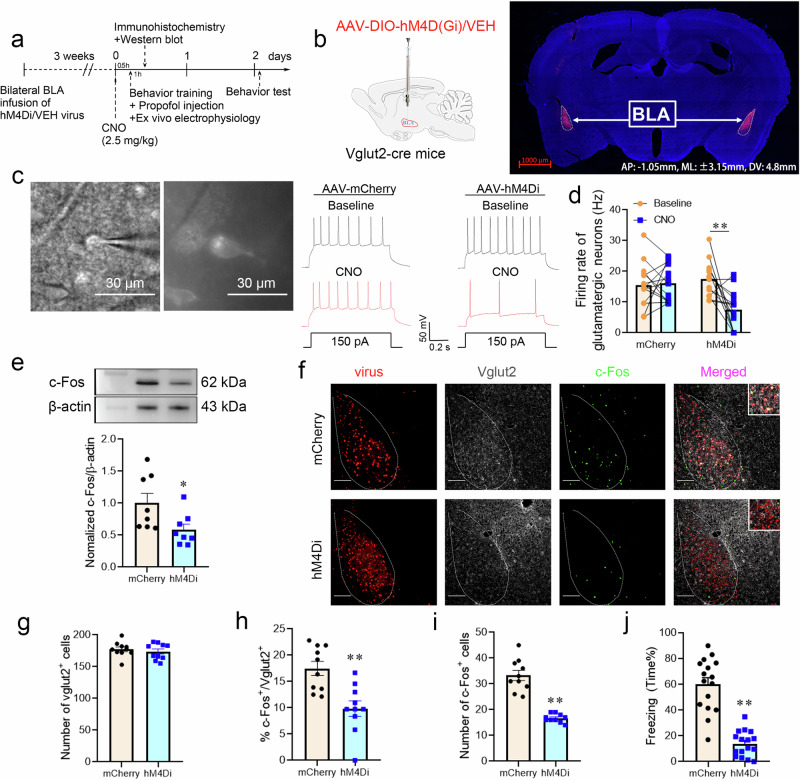
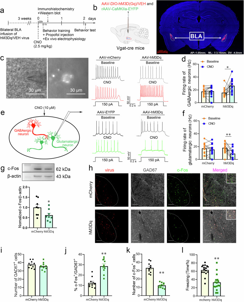
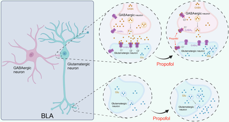
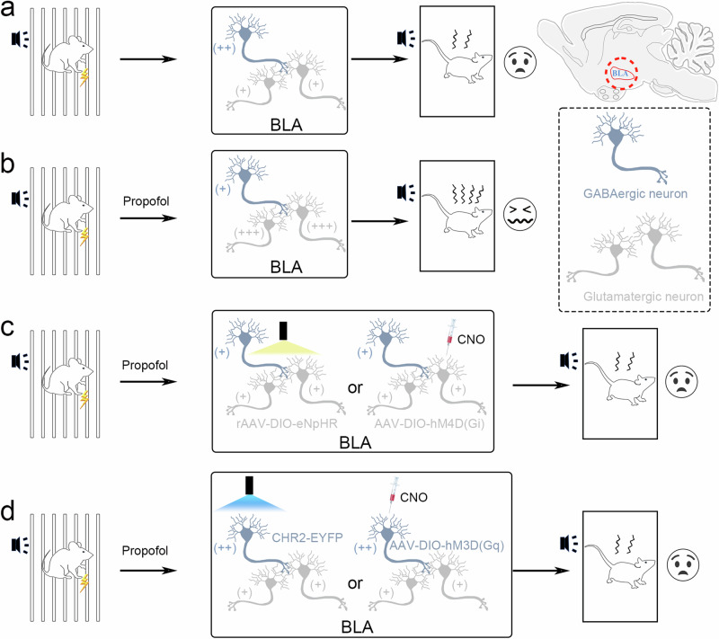
Posttraumatic stress disorder (PTSD) is associated with glutamatergic neuron hyperactivation in the basolateral amygdala (BLA) brain area, while GABAergic interneurons in the BLA modulate glutamatergic neuron excitability. Studies have shown that propofol exerts its effects through potentiation of the inhibitory neurotransmitter γ-aminobutyric acid. The neuronal mechanism by which propofol anesthesia modulates fear memory is currently unknown. Here, we used optogenetics and chemogenetics to suppress glutamatergic neurons or activate GABAergic interneurons in the BLA to assess alterations in neuronal excitation-inhibition balance and investigate fear memory. The excitability of glutamatergic neurons in the BLA was significantly reduced by the suppression of glutamatergic neurons or activation of GABAergic interneurons, while propofol-mediated enhancement of fear memory was attenuated. We suggest that propofol anesthesia could reduce the excitability of GABAergic neurons through activation of GABAA receptors, subsequently increasing the excitability of glutamatergic neurons in the mice BLA; the effect of propofol on enhancing mice fear memory might be mediated by strengthening glutamatergic neuronal excitability and decreasing the excitability of GABAergic neurons in the BLA; neuronal excitation-inhibition imbalance in the BLA might be important in mediating the enhancement of fear memory induced by propofol.


Conflict of interest statement

The authors declare no competing interests.

Figures

Fig. 1
Fig. 1. Fear memory was enhanced by propofol after FC training in mice.
a Diagram of fear conditioning (FC) training and fear conditioning test (FCT) patterns. b Freezing in response to the conditioned tone after the infusion of different doses of propofol. Propofol enhanced freezing at doses of 60 × 4 and 60 × 5 mg/kg but not 60 × 2 and 60 × 3 mg/kg (n = 16/group, one-way ANOVA, F (4, 75) = 11.66, p < 0.001, **p < 0.01). c Freezing in response to the conditioned tone after the infusion of vehicle or propofol at different time points after the conditioning. Propofol enhanced freezing at 0 and 30 min but not at 60 min or 90 min (n = 16/group, two-tailed unpaired t test, **p < 0.01). d Representative images of Western blots and quantification of c-Fos expression among the 4 groups (n = 10, one-way ANOVA, F (3, 36) = 18.82, p < 0.0001, *p = 0.030, **p < 0.01). e Representative images of c-Fos/DAPI immunofluorescence in BLA neurons after vehicle or propofol treatment; scale bar, 100 µm. f The number of c-Fos+ cells in the BLA in mice that were administered propofol after FC training was increased compared to that in trained mice administered the vehicle (n = 20/group, one-way ANOVA, F (3, 76) = 349.8, p < 0.0001, **p < 0.01). g Freezing was comparable across all groups during FC training (n = 16/group, Kruskal-Wallis test, p = 0.523). h Trained mice administered propofol showed enhanced fear freezing compared to those administered the vehicle (n = 16/group, one-way ANOVA, F (3, 60) = 75.48, p < 0.001, **p < 0.01). All data are presented as the mean ± SEM.
Fig. 2
Fig. 2. The excitability of glutamatergic and GABAergic neurons in the BLA was enhanced or attenuated, respectively, by propofol anesthesia after FC training in mice.
a Representative images of c-Fos/DAPI/Vglut2 immunofluorescence in BLA neurons after vehicle or propofol treatment; scale bar, 100 µm. b No significant difference was detected in the percentage of Vglut2+ cells between mice administered vehicle or propofol (n = 10/group, two-tailed unpaired t test). c The percentage of c-Fos+ & Vglut2+ cells in the BLA in trained mice administered propofol was higher than that in trained mice administered vehicle (n = 10 mice/group, Mann–Whitney U test, **p < 0.01). d Representative images of c-Fos/DAPI/GAD67 immunofluorescence in BLA neurons after vehicle or propofol treatment; scale bar, 100 µm. e No significant difference was detected in the percentage of GAD67+ cells between mice administered vehicle or propofol (n = 10/group, two-tailed unpaired t test). f The percentage of c-Fos+& GAD67+ cells in the BLA in trained mice administered propofol was decreased compared to that in trained mice administered vehicle (n = 10 mice/group, two-tailed unpaired t test, **p < 0.01). All data are presented as the mean ± SEM.
Fig. 3
Fig. 3. The excitability of glutamatergic and GABAergic neurons in the BLA was enhanced or attenuated, respectively, by propofol anesthesia after FC training in mice.
a The electrophysiology of GABAergic (mCherry+) neurons in the BLA of Vgat-cre mice. A typical trace of membrane potential observed before and after the application of propofol (5 µM). Firing rate of action potentials evoked by depolarizing current pulses of 0–200 pA, values were presented (n = 13 neurons from 6 mice, two-way repeated-measures ANOVA, F (20, 504) = 2.936, p < 0.0001, **p < 0.01 vs. Control group). Threshold currents that evoked the first action potential (b) and comparison of resting membrane potential (c), (n = 13 neurons from 6 mice, two-tailed paired t test and two-tailed Wilcoxon matched-pairs signed rank test, **p < 0.01). d The electrophysiology of glutamatergic neurons (mCherry+) in the BLA of Vglut2-cre mice. A typical trace of membrane potential observed before and after the application of propofol (5 µM). Firing rate of action potentials evoked by depolarizing current pulses of 0–200 pA, values were presented (n = 13 neurons from 6 mice, two-way repeated-measures ANOVA, F (20, 502) = 5.073, p < 0.0001, **p < 0.01). e, f Threshold currents that evoked the first action potential and comparison of resting membrane potential, (n = 13 neurons from 6 mice, two-tailed Wilcoxon matched-pairs signed rank test, **p < 0.01). g The electrophysiology of GABAergic neurons (mCherry+) perfused bicuculline (30 µM) in the BLA of Vgat-cre mice. A typical trace of membrane potential observed before and after the application of propofol (5 µM). Firing rate of action potentials evoked by depolarizing current pulses of 0–200 pA, values were presented (n = 13 neurons from 6 mice, two-way repeated-measures ANOVA, F (20, 504) = 0.076, p > 0.999). h Threshold currents that evoked the first action potential, values were presented (n = 13 neurons from 6 mice, two-tailed Wilcoxon matched-pairs signed rank test, p > 0.05). i Comparison of resting membrane potential (n = 13 neurons from 6 mice, two-tailed paired t test, p > 0.05). j The electrophysiology of glutamatergic neurons (mCherry+) perfused bicuculline (30 µM) in the BLA of Vglut2-cre mice. A typical trace of membrane potential observed before and after the application of propofol (5 µM). Firing rate of action potentials evoked by depolarizing current pulses of 0–200 pA, values were presented (n = 13 neurons from 6 mice, two-way repeated-measures ANOVA, F (20, 504) = 0.212, p > 0.999). k Threshold currents that evoked the first action potential, values were presented (n = 13 neurons from 6 mice, two-tailed paired t test, p > 0.05). l Comparison of resting membrane potential (n = 13 neurons from 6 mice, two-tailed Wilcoxon matched-pairs signed rank test, p > 0.05). m Representative traces of sEPSC from glutamatergic neurons (mCherry+) in the BLA of Vglut2-cre mice. Scale bars = 50 pA, 10 s. n The sEPSC frequency in glutamatergic neurons was significantly increased in brain slices perfused propofol (5 µM) compared with those from control (n = 7 neurons from 6 mice, two-tailed paired t test, *p = 0.032). While The sEPSC frequency showed no statistical difference in brain slices perfused bicuculline + propofol and slices perfused bicuculline (n = 6 neurons from 6 mice, two-tailed paired t test, p > 0.05). o The sEPSC amplitude in glutamatergic neurons was significantly increased in brain slices perfused propofol (5 µM) compared with those from control (n = 7 neurons from 6 mice, two-tailed paired t test, *p = 0.013). While The sEPSC amplitude showed no statistical difference in brain slices perfused bicuculline + propofol and slices perfused bicuculline (n = 6 neurons from 6 mice, two-tailed paired t test, p > 0.05). All data are presented as the mean ± SEM.
Fig. 4
Fig. 4. Inhibiting glutamatergic neurons in the BLA by optogenetic regulation attenuated the enhanced effect of propofol on fear memory.
a Experimental time course for surgery, FC training, propofol injection, photoinhibition, and FCT. b Schematic of 2/9rAAV-DIO-eNpHR-mCherry or 2/9rAAV-DIO-mCherry injection and optic fiber implantation into the BLA of Vglut2-cre mice; representative immunohistochemical staining (scale bar, 1000 µm, AP: -1.05 mm, ML: ±3.15 mm, DV: 4.8 mm); schematic of fiber placement and viral spread in the BLA (scale bar, 100 µm, AP: -1.05 mm). c Neurons expressing mCherry fluorescence were observed in the BLA when ex vivo brain slices were exposed under a microscope. The pulse train evokes outward currents and abolishes action potentials (30 pA) in glutamatergic neurons. d Representative Western blot image and quantification of c-Fos expression. The expression of c-Fos was reduced after the photoinhibition of glutamatergic neurons in the BLA (n = 8, two-tailed unpaired t test, *p = 0.012). e Representative images of mCherry/Vglut2/c-Fos immunofluorescence in BLA neurons after virus treatment and photoinhibition; scale bar, 100 µm. fh The ratio of c-Fos+ & Vglut2+ cells and the total number of c-Fos+ cells in the BLA after the photoinhibition of glutamatergic neurons were reduced (n = 10/group, two-tailed unpaired t test, **p < 0.01). i Reduction in freezing in mice after the photoinhibition of glutamatergic neurons in the BLA (n = 16/group, two-tailed unpaired t test, **p < 0.01). All data are presented as the mean ± SEM.
Fig. 5
Fig. 5. Activating GABAergic interneurons in the BLA by optogenetic regulation attenuated the enhanced effect of propofol on fear memory.
a Experimental time course for surgery, FC training, propofol injection, photoactivation, and FCT. b Schematic of fiber optic implantation and rAAv-CaMKIIa-mCherry injection into the BLA of Vgat-ChR2-EYFP mice. Schematic of fiber optic implantation into the BLA of Vgat-ChR2-EYFP mice. c Neurons expressing EYFP fluorescence were observed in the BLA when ex vivo brain slices were exposed under a microscope. d Trains of pulses (pulses at 5, 10, 15, and 20 Hz) evoke reproducible currents (top) and action potentials (bottom) in a GABAergic interneuron. e Light illumination was applied to the BLA slice, and the activity of glutamatergic neurons was recorded. f Photoactivation (20 Hz) of GABAergic interneurons reduced firing in glutamatergic neurons, and firing recovered after the light illumination was terminated. g Representative Western blot image and quantification of c-Fos expression. The expression of c-Fos was reduced after photoactivation of GABAergic interneurons in the BLA (n = 8, two-tailed unpaired t test, **p < 0.01). h Representative images of DAPI/GAD67/c-Fos immunofluorescence in BLA neurons after photoactivation; scale bar, 100 µm. The ratio of c-Fos+ & GAD67+ cells in the BLA was increased after photoactivation of GABAergic interneurons, while the total number of c-Fos+ cells was decreased (n = 10/group), Mann–Whitney U test (i, j), two-tailed unpaired t test (k), *p = 0.01, **p < 0.01. l Reduction in freezing in mice after photoactivation of GABAergic interneurons in the BLA (n = 16/group, two-tailed unpaired t test, **p < 0.01). All data are presented as the mean ± SEM.
Fig. 6
Fig. 6. Inhibiting glutamatergic neurons in the BLA by chemogenetic regulation attenuated the effect of propofol on enhancing fear memory.
a Experimental time course for surgery, CNO injection, FC training, propofol injection, and FCT. b Schematic of 2/9AAV-DIO-hM4D(Gi)-mCherry or 2/9AAV-DIO-mCherry injection into the BLA of Vglut2-cre mice. Representative immunohistochemical staining (scale bar, 1000 µm, AP: -1.05 mm, ML: ±3.15 mm, DV: 4.8 mm). c Neurons expressing mCherry fluorescence were observed in the BLA when ex vivo brain slices were exposed under a microscope. Bath application of CNO (10 µM) decreased the firing rate in hM4Di-positive BLA neurons in vitro. d Quantification of the firing rate (n = 14 cells from 6 mice injected with AAV-VEH and n = 14 cells from 6 mice injected with AAV-hM4Di, two-tailed paired t test, **p < 0.01). e Representative Western blot image and quantification of c-Fos expression. The expression of c-Fos was reduced after chemogenetic inhibition of glutamatergic neurons in the BLA (n = 8, two-tailed unpaired t test, *p = 0.033). f Representative images of mCherry/Vglut2/c-Fos immunofluorescence in BLA neurons after virus treatment and chemogenetic manipulation; scale bar, 100 µm. gi The percentage of c-Fos+ & Vglut2+ cells and the total number of c-Fos+ cells were reduced after chemogenetic inhibition of glutamatergic neurons in the BLA (n = 10/group), two-tailed unpaired t test, **p < 0.01. j Reduction in freezing in mice after chemogenetic inhibition of glutamatergic neurons in the BLA (n = 16/group, two-tailed unpaired t test, **p < 0.01). All data are presented as the mean ± SEM.
Fig. 7
Fig. 7. Activating GABAergic interneurons in the BLA by chemogenetic regulation attenuated the effect of propofol on enhancing fear memory.
a Experimental time course for surgery, CNO injection, FC training, propofol injection, and FCT. b Schematic of 2/9AAV-DIO-hM3D(Gq)-mCherry or 2/9AAV-DIO-mCherry and rAAV-CaMKIIa-EYFP injection into the BLA of Vgat-cre mice. Representative immunohistochemical staining (scale bar, 1000 µm, AP: -1.05 mm, ML: ±3.15 mm, DV: 4.8 mm). c Neurons expressing mCherry fluorescence were observed in the BLA when in vitro brain slices were exposed under a microscope. Bath application of CNO (10 µM) increased the firing rate in hM3Dq-positive BLA neurons in vitro. d Quantification of the firing rate (n = 14 cells from 6 mice injected with AAV-VEH and n = 14 cells from 6 mice injected with AAV-hM3Dq, two-tailed Wilcoxon matched-pairs signed rank test, *p = 0.013). e Chemogenetic activation was applied to the BLA slice, and the activity of glutamatergic neurons was recorded. Bath application of CNO (10 µM) decreased the firing rate in BLA glutamatergic neurons in vitro. f Quantification of the firing rate (n = 12 cells from 6 mice injected with AAV-VEH+ rAAV-CaMKIIa-EYFP and n = 13 cells from 6 mice injected with AAV-hM3Dq+ rAAV-CaMKIIa-EYFP, two-tailed paired t test, **p < 0.01). g Representative Western blot image and quantification of c-Fos expression. The expression of c-Fos was reduced after chemogenetic activation of GABAergic interneurons in the BLA (n = 8, two-tailed unpaired t test, *p = 0.045). h Representative images of mCherry/GAD67/c-Fos immunofluorescence in BLA neurons after virus treatment and chemogenetic manipulation; scale bar, 100 µm. Ik The ratio of c-Fos+ & GAD67+ cells in the BLA was increased after chemogenetic activation of GABAergic interneurons, while the total number of c-Fos+ cells was decreased (n = 10/group, two-tailed unpaired t test, **p < 0.01). l Reduction in freezing in mice after chemogenetic activation of GABAergic interneurons in the BLA (n = 16/group, two-tailed unpaired t test, **p < 0.01). All data are presented as the mean ± SEM.
Fig. 8
Fig. 8. The disinhibitory effect of GABAergic neurons on glutamatergic neurons is stronger than the potentiation of GABAA receptors on glutamatergic neurons by propofol itself, leading to the manifestation of activating effects on glutamatergic neurons.
Created in BioRender. Ning, W. (2023) BioRender.com/k92f213.
Fig. 9
Fig. 9. Inhibiting glutamatergic neurons or activating GABAergic interneurons in the BLA attenuated the effect of propofol on enhancing fear memory.
a, b Fear memory was enhanced by propofol after FC training in mice. c Inhibiting glutamatergic neuron activity in the BLA attenuated the effect of propofol on enhancing fear memory. d Activating GABAergic interneurons in the BLA attenuated the effect of propofol on enhancing fear memory.

References

    1. Harnett, N., Goodman, A. & Knight, D. PTSD-related neuroimaging abnormalities in brain function, structure, and biochemistry. Exp. Neurol.330, 113331 (2020). - PubMed
    1. Brewin, C. et al. A review of current evidence regarding the ICD-11 proposals for diagnosing PTSD and complex PTSD. Clin. Psychol. Rev.58, 1–15 (2017). - PubMed
    1. Bisson, J., Cosgrove, S., Lewis, C. & Robert, N. Post-traumatic stress disorder. BMJ351, h6161 (2015). - PMC - PubMed
    1. Rytwinski, N., Scur, M., Feeny, N. & Youngstrom, E. The co-occurrence of major depressive disorder among individuals with posttraumatic stress disorder: a meta-analysis. J. Trauma. stress26, 299–309 (2013). - PubMed
    1. Cougle, J., Resnick, H. & Kilpatrick, D. PTSD, depression, and their comorbidity in relation to suicidality: cross-sectional and prospective analyses of a national probability sample of women. Depression Anxiety26, 1151–1157 (2009). - PubMed

LinkOut - more resources