Review
. 2025 Jan 2;26(1):e202400863.
doi: 10.1002/cphc.202400863. Epub 2024 Nov 20.

Radiation and DNA Origami Nanotechnology: Probing Structural Integrity at the Nanoscale

Affiliations
Review

Radiation and DNA Origami Nanotechnology: Probing Structural Integrity at the Nanoscale

João Ameixa et al. Chemphyschem. .

Abstract

DNA nanotechnology has emerged as a groundbreaking field, using DNA as a scaffold to create nanostructures with customizable properties. These DNA nanostructures hold potential across various domains, from biomedicine to studying ionizing radiation-matter interactions at the nanoscale. This review explores how the various types of radiation, covering a spectrum from electrons and photons at sub-excitation energies to ion beams with high-linear energy transfer influence the structural integrity of DNA origami nanostructures. We discuss both direct effects and those mediated by secondary species like low-energy electrons (LEEs) and reactive oxygen species (ROS). Further we discuss the possibilities for applying radiation in modulating and controlling structural changes. Based on experimental insights, we identify current challenges in characterizing the responses of DNA nanostructures to radiation and outline further areas for investigation. This review not only clarifies the complex dynamics between ionizing radiation and DNA origami but also suggests new strategies for designing DNA nanostructures optimized for applications exposed to various qualities of ionizing radiation and their resulting byproducts.

Keywords: DNA damage; DNA structures; Nanostructures; Nanotechnology.


Conflict of interest statement

The authors declare no conflict of interest.

Figures

Figure 1
Figure 1
Energy spectrum illustrating the range of radiation types discussed in this review, ranging from 10−1–1010 eV, and their interactions with DNA. Highlighted are photons (blue) including UV (A, B, C), VUV, XUV, high‐energy photons, and γ‐rays; electrons (green) encompassing low‐energy electrons (LEEs, <20 eV), high‐energy electrons, and β particles. Ion beams are shown in the bottom‐most block with nuclear stopping mechanisms typically dominating at the lower energy range (as highlighted in red) and electronic stopping mechanisms at high energy (in dark blue), although these ranges vary depending on the energy, size, and charge state of the ion projectile.
Figure 2
Figure 2
(A) A summary of commonly observed UV‐induced structural damages and modifications to DNA origami nanostructures spanning the UVA‐UVC range. (B.I) AFM images of the control sample (a) and UVA‐irradiated DNA nanotriangles loaded with BMEPC which acts as a photosensitizer. Adapted with permission from Ref. [65] Copyright 2016 American Chemical Society. (b). (B.II) AFM scans in liquid of plain origami tiles annealed to 60 °C (a) and those which were cross‐linked by 8‐methoxypsoralen (8‐MOP) at proximal thymine residues upon activation by UVA light and then also annealed to 60 °C (b), demonstrating enhanced thermal stability by cross‐linking. Adapted with permission from Ref. [67] Copyright 2011 American Chemical Society. (B.III) Electrophoresis results of UVB cross‐linked and uncross‐linked DNA origami bundles heated to different temperatures. The bundles contain strategically placed proximal thymidine residues that link into CPDs upon UVB irradiation. Reprinted with permission from Ref. [27] Copyright 2018 American Association for the Advancement of Science. An average 2D particle TEM micrograph of the CPD‐cross‐linked sample in double‐distilled water is shown on the rightmost image. (B. IV) AFM images of DNA origami nanoribbons formed from rectangular DNA origami tiles before (a) and after mild irradiation using UVC light showing induced conformational changes that take advantage of the stress relaxation from the nicking of DNA strands by (b) UVC. Adapted with permission from Ref. [64] Copyright 2024 American Chemical Society.
Figure 3
Figure 3
(A) A scheme of typical damages caused by low to medium LET radiation such as γ‐rays and proton beams to 2D DNA origami nanotriangles in solution. The single‐stranded domains on the corners of the 2D DNA origami triangle are compromised and although the overall structure is preserved, unraveling the structure in electrophoresis show absorbed‐dose‐dependent strand breaking in the origami scaffold. Reproduced from Ref. [85] with permission from the Royal Society of Chemistry. (B) A summary of typical damages on 2D DNA origami triangles in dry conditions on Si substrates irradiated by low to medium LET radiation (I) and high‐LET radiation in the form of ion beams (II) presenting opportunities for localized nanoscale processing of DNA origami nanostructures. Adapted with permission from Ref. [93]
Figure 4
Figure 4
(A) Schematic outlining the experimental procedure for absolute quantification of DNA single‐strand breaks induced by radiation. Adapted with permission from Ref. [11] Copyright 2024 American Chemical Society. (B) AFM images comparing (I.) a control sample, and (II.) samples exposed to 10 eV LEEs under high‐vacuum conditions for 40 seconds. Adapted with permission from Ref. [99] Copyright 2017 Springer Nature. (C) Effects of 8‐bromoadenine (8BrA) modifications on DNA sequences, namely: (I.) low‐energy electron‐induced single strand breaks in sequences TT(XTA)3TT, where X=8BrA, A, and (II.) comparison of obtained EFSB as a function of the electron energy along with the average EFSB(8BrA). Adapted with permission from Ref. [98] Copyright 2017 Wiley‐VCH.
Figure 5
Figure 5
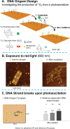
(A) Schematic representation of the experimental approach for studying 1O2 production and diffusion from a centrally positioned photosensitizer (IPS) towards SOC linkers upon exposure to red light. (B) AFM images comparing (1) a non‐irradiated control sample and (2) a sample after irradiation, showing the effects of photoexcitation. (C) Distance‐dependent analysis using (1) a DNA origami structure with DNA groups placed at three distinct positions and (2) the relative DNA strand breakage upon photoexcitation as a function of the distance from the IPS. Adapted with permission from Ref. [102] Copyright 2010 American Chemical Society.
Figure 6
Figure 6
(A) Schematic of the experimental procedure for the determination of absolute DNA strand break cross sections, σSB , upon photoexcitation of the water‐soluble photosensitizer [Cr(ddpd)2][BF4]3.(B) AFM images depicting a) a DNA origami control sample kept in the dark and b) a DNA origami sample treated with the photosensitizer and exposed to UV light (365 nm, 0.60 mWcm−2) for 120 seconds. Insets show the results before and after exposure. (C) 1. Absolute DNA strand break cross sections for ssDNA (black) and dsDNA (red) and 2. Quantum yields as function of the photosensitizer's concentration. Adapted with permission from Ref. [104] Copyright 2023 Wiley‐VCH.

References

    1. Rothemund P. W. K., Nature 2006, 440, 297–302. - PubMed
    1. Tapio K., Bald I., Multifunct. Mater. 2020, 3, 032001.
    1. Engelhardt F. A. S., Praetorius F., Wachauf C. H., Brüggenthies G., Kohler F., Kick B., Kadletz K. L., Pham P. N., Behler K. L., Gerling T., Dietz H., ACS Nano 2019, 13, 5015–5027. - PMC - PubMed
    1. Dey S., Fan C., Gothelf K. V., Li J., Lin C., Liu L., Liu N., Nijenhuis M. A. D., Saccà B., Simmel F. C., Yan H., Zhan P., Nat. Rev. Methods Primers 2021, 1, 1–24.
    1. Hong F., Zhang F., Liu Y., Yan H., Chem Rev 2017, 117, 12584–12640. - PubMed

LinkOut - more resources