

Isolated melanoma metastasis in a patient with large congenital nevus without detectable primary melanoma: a case report and review of literature
- PMID: 39473498
- PMCID: PMC11518708
- DOI: 10.3389/fmed.2024.1427982
Isolated melanoma metastasis in a patient with large congenital nevus without detectable primary melanoma: a case report and review of literature
Abstract
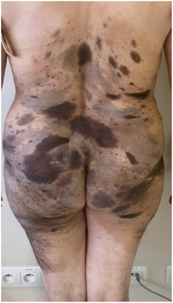
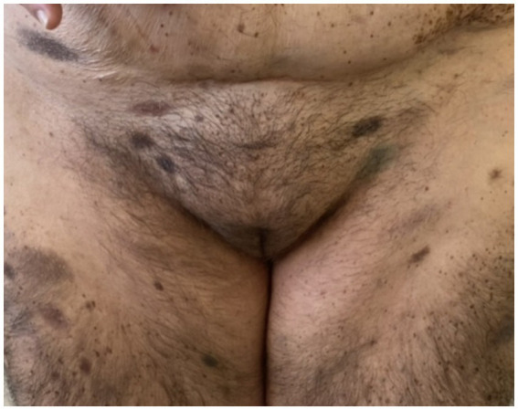
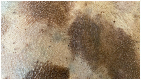
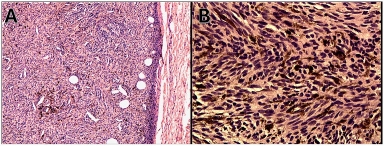
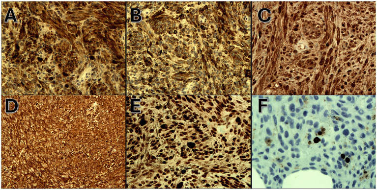
Giant congenital pigmented nevi constitute an extremely diverse group of skin lesions with varying morphologies. These nevi are often associated with many clinical implications, such as increased risk of melanoma and the presence of neurocutaneous melanosis, with melanoma being the primary concern. We present a rare case of a 62-year-old patient with a giant congenital birthmark who reported to the oncology department due to a tumor in the lower abdomen detected during an ultrasound examination. A biopsy of the lesion showed the presence of melanoma metastasis. Four independent dermatologists performed a dermoscopic examination of the patient's skin and mucous membranes. In the PET/CT examination, apart from the previously described change in the lower abdomen, no metabolically active foci with features of malignant growth were found. The patient underwent surgical removal of the lesion in the lower abdomen. The postoperative histopathological examination confirmed the presence of metastasis of melanoma in the subcutaneous tissue of the abdomen with no connection to the epidermis. The BRAFV600 mutation was not found in the molecular test. For stage IV R0 melanoma with distant metastasis, with stage T0N0M1a, the only adjuvant treatment option following radical resection is nivolumab. After a rheumatological consultation, the patient was qualified for adjuvant treatment with nivolumab.
Keywords: adjuvant treatment; giant congenital melanocytic nevus; melanoma; melanoma of unknown primary origin; primary melanoma.
Copyright © 2024 Pabianek, Jatczak-Grochala, Lesiak, Narbutt, Siekierko, Stasikowska-Kanicka and Ciążyńska.
Conflict of interest statement
The authors declare that the research was conducted in the absence of any commercial or financial relationships that could be construed as a potential conflict of interest.
Figures
Similar articles
-
Role of In Vivo Reflectance Confocal Microscopy in the Analysis of Melanocytic Lesions.Acta Dermatovenerol Croat. 2018 Apr;26(1):64-67. Acta Dermatovenerol Croat. 2018. PMID: 29782304 Review.
-
Giant lung metastasis of NRAS-mutant melanoma in a 24-year-old patient with a history of BRAF-mutant conventional melanoma harboring Spitzoid morphology: a case report.Diagn Pathol. 2020 Oct 25;15(1):132. doi: 10.1186/s13000-020-01046-3. Diagn Pathol. 2020. PMID: 33100226 Free PMC article.
-
Melanoma Developing from an Intradermal Nevus: Report on Two Patients.Acta Dermatovenerol Croat. 2023 Aug;31(1):40-42. Acta Dermatovenerol Croat. 2023. PMID: 37843090
-
The risk of melanoma and neurocutaneous melanosis associated with congenital melanocytic nevi.Semin Cutan Med Surg. 2010 Sep;29(3):159-64. doi: 10.1016/j.sder.2010.06.007. Semin Cutan Med Surg. 2010. PMID: 21051009 Review.
-
Giant congenital melanocytic nevi: the significance of neurocutaneous melanosis in neurologically asymptomatic children.Plast Reconstr Surg. 2001 Apr 1;107(4):933-41. doi: 10.1097/00006534-200104010-00005. Plast Reconstr Surg. 2001. PMID: 11252085
References
-
- World Health Organization International agency for research on cancer (IARC) GLOBOCAN. (2020). Available at: https://gco.iarc.fr/today/home (Accessed April 14, 2024).
Publication types
LinkOut - more resources
Full Text Sources