

Cancer and lymphatic marker FOXC2 drives wound healing and fibrotic tissue formation
- PMID: 39473610
- PMCID: PMC11518795
- DOI: 10.3389/fphys.2024.1427113
Cancer and lymphatic marker FOXC2 drives wound healing and fibrotic tissue formation
Abstract
Introduction: The FOXC2 transcription factor has been tied to a wide range of disease states, serving as a promising prognostic biomarker associated with aggressive basal-like human breast cancers (increased cancer invasion and metastasis). Dysregulation of FOXC2 expression has also been found to promote defects in lymphatic remodeling and hyperplastic lymphedema-distichiasis (LD). Since chronic lymphedema is a forerunner of several malignancies and cancers have been known to arise from poorly healing chronic wounds (e.g., Marjolin ulcers), we examined the effect of Foxc2 dysfunction on skin wound healing.
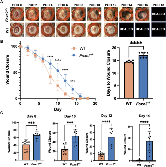
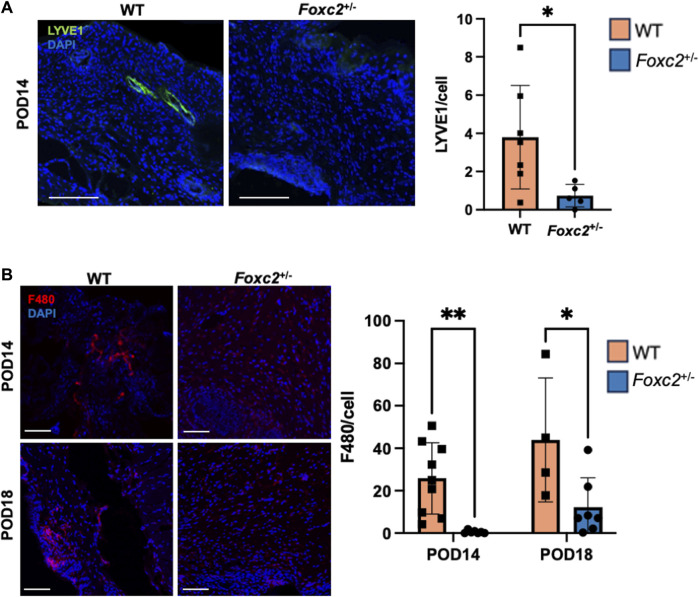
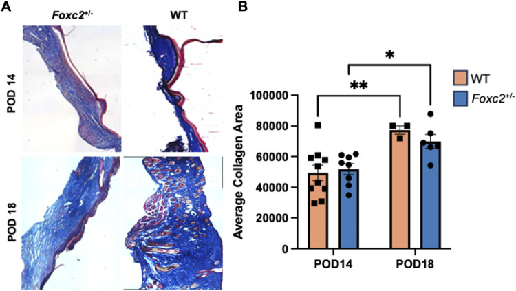
Methods: We used our splinted excisional wounding model that mimics human-like wound healing on wildtype and Foxc2+/- mice (n = 4), which demonstrate incomplete lymphatic vasculature and lymphatic dysfunction. Wound size was measured over the course of 18 days. Tissue was explanted from both groups at post-operative day (POD) 14 and 18 and stained with Masson's Trichrome to assess scar formation, Picrosirius Red for dermal integrity, or immunofluorescence to assess lymphatic (LYVE1) cell populations.
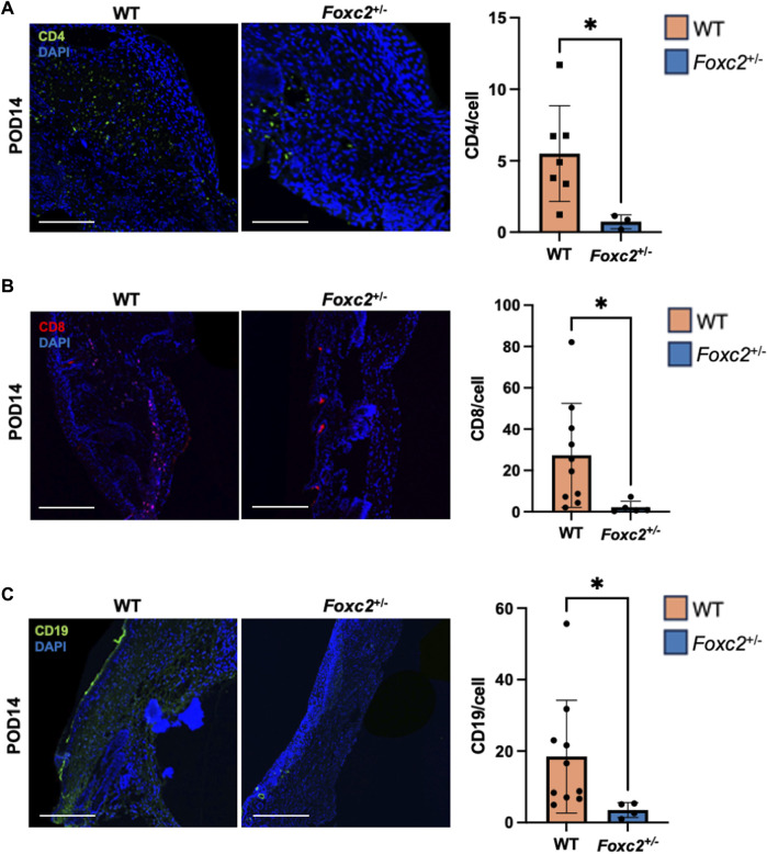
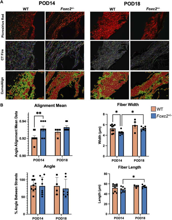
Results: Wildtype mice completely healed by POD 14, while Foxc2+/-mice did not completely heal until POD18. Scar area of healed Foxc2+/-mice (POD 18) was larger than that of healed wild-type mice (POD 14; p = 0.0294). At POD 14, collagen "bers in the scars of Foxc2+/-mice to be narrower (p = 0.0117) and more highly aligned (p = 0.0110), indicating signi"cantly more "brosis in these mice. Collagen "bers in both groups became longer (p = 0.0116) and wider (p = 0.0020) from POD 14 to 18, indicating a temporal evolution of "brosis. Foxc2+/-mice also had lower numbers of LYVE1+, F4/80+ and CD4+ cells compared to wildtype mice.
Discussion: Individuals over 65 years old are more likely to develop cancer and are highly susceptible to developing chronic wounds. Here, we found that FOXC2, which is tied to cancer metastasis and lymphatic dysregulation, also impairs wound healing and promotes "brotic tissue architecture. With FOXC2 proposed as a potential therapeutic target for cancer metastasis, its downstream systemic effects should be considered against the increased chance of developing nonhealing wounds. Further delineation of the microenvironment, cellular events, and molecular signals during normal and Foxc2-associated abnormal wound healing will improve clinical therapies targeting this important marker.
Keywords: FOXC2; fibrosis; inflammation; lymphatics; wound healing.
Copyright © 2024 Granoski, Fischer, Hahn, Sivaraj, Kussie, Quintero, Alsharif, McKenna, Yasmeh, Hostler, Mora Pinos, Erickson, Witte, Chen, Gurtner.
Conflict of interest statement
The authors declare that the research was conducted in the absence of any commercial or financial relationships that could be construed as a potential conflict of interest.
Figures
Similar articles
-
[Effect of hydrocinnamoyl-L-valyl pyrrolidine on healing quality of deep partial-thickness scald wound in mice].Zhonghua Shao Shang Za Zhi. 2016 Nov 20;32(11):658-666. doi: 10.3760/cma.j.issn.1009-2587.2016.11.005. Zhonghua Shao Shang Za Zhi. 2016. PMID: 27894387 Chinese.
-
[Influence of porcine urinary bladder matrix and porcine acellular dermal matrix on wound healing of full-thickness skin defect in diabetic mice].Zhonghua Shao Shang Za Zhi. 2020 Dec 20;36(12):1130-1138. doi: 10.3760/cma.j.cn501120-20200901-00399. Zhonghua Shao Shang Za Zhi. 2020. PMID: 33379849 Chinese.
-
Avenanthramide and β-Glucan Therapeutics Accelerate Wound Healing Via Distinct and Nonoverlapping Mechanisms.Adv Wound Care (New Rochelle). 2024 Apr;13(4):155-166. doi: 10.1089/wound.2023.0050. Epub 2024 Feb 26. Adv Wound Care (New Rochelle). 2024. PMID: 38299969
-
Scar-free healing: from embryonic mechanisms to adult therapeutic intervention.Philos Trans R Soc Lond B Biol Sci. 2004 May 29;359(1445):839-50. doi: 10.1098/rstb.2004.1475. Philos Trans R Soc Lond B Biol Sci. 2004. PMID: 15293811 Free PMC article. Review.
-
Lymphangiogenesis: novel strategies to promote cutaneous wound healing.Burns Trauma. 2024 Sep 26;12:tkae040. doi: 10.1093/burnst/tkae040. eCollection 2024. Burns Trauma. 2024. PMID: 39328366 Free PMC article. Review.
References
LinkOut - more resources
Full Text Sources
Research Materials
Miscellaneous