Review
. 2023 Aug 2;2(2):81-97.
doi: 10.1021/cbmi.3c00070. eCollection 2024 Feb 26.

Advances in Small-Molecule Fluorescent pH Probes for Monitoring Mitophagy

Affiliations
Review

Advances in Small-Molecule Fluorescent pH Probes for Monitoring Mitophagy

Yurui Liu et al. Chem Biomed Imaging. .

Abstract

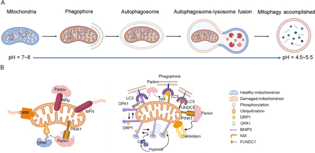
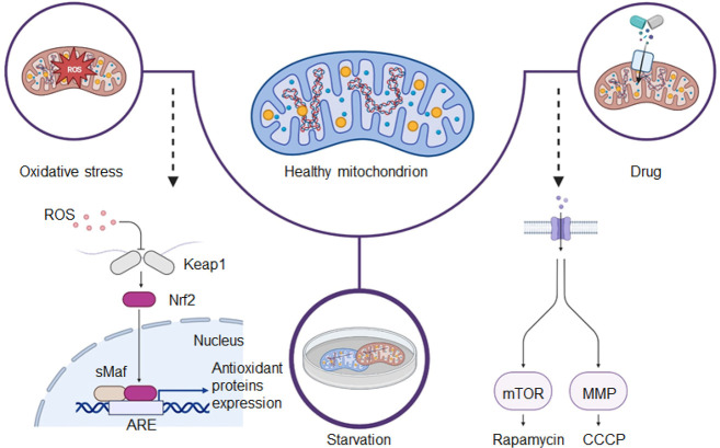
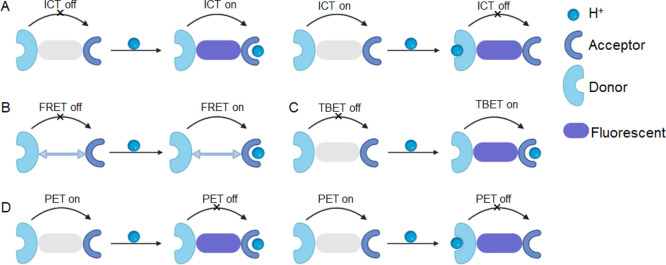
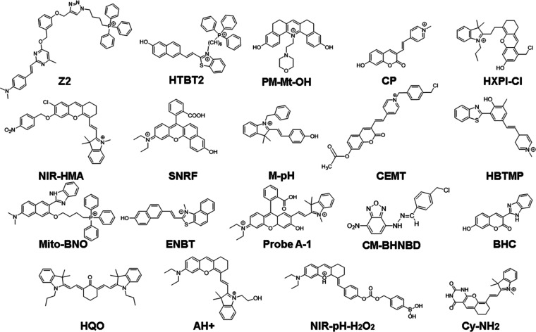
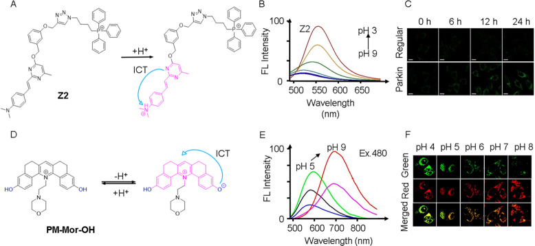
Mitochondria play a crucial role in regulating cellular energy homeostasis and cell death, making them essential organelles. Maintaining proper cellular functions relies on the removal of damaged mitochondria through a process called mitophagy. Mitophagy is associated with changes in the pH value and has implications for numerous diseases. To effectively monitor mitophagy, fluorescent probes that exhibit high selectivity and sensitivity based on pH detection have emerged as powerful tools. In this review, we present recent advancements in the monitoring of mitophagy using small-molecule fluorescence pH probes. We focus on various sensing mechanisms employed by these probes, including intramolecular charge transfer (ICT), fluorescence resonance energy transfer (FRET), through bond energy transfer (TBET), and photoelectron transfer (PET). Additionally, we discuss disease models used for studying mitophagy and summarize the design requirements for small-molecule fluorescent pH probes suitable for monitoring the mitophagy process. Lastly, we highlight the remaining challenges in this field and propose potential directions for the future development of mitophagy probes.


Conflict of interest statement

The authors declare no competing financial interest.

Figures

Figure 1
Figure 1
(A) Mitophagy process. (B) PINK1 and E3 Parkin-mediated pathway (left) and outer-membrane mitophagy receptor-mediated pathway (right).
Figure 2
Figure 2
Illustration of three pathways used to construct mitophagy models.
Figure 3
Figure 3
Schematic representation of the fluorescent mechanisms discussed in this review. (A) Intramolecular charge transfer (ICT). (B) Fluorescence resonance energy transfer (FRET). (C) Through-bond energy transfer (TBET). (D) Photoinduced electron transfer (PET).
Figure 4
Figure 4
Small-molecule fluorescent pH probes based on the ICT mechanism.
Figure 5
Figure 5
(A) Molecular structure and pH sensing mechanism of probe Z2. (B) Fluorescence spectra of Z2 (λex = 450 nm). (C) HeLa cells and Parkin-HeLa cells pretreated with 10 μM CCCP, and then stained with Z2. λex/em = 458/480–520 nm. Scale bar = 20 μm. (A–C) Reproduced with permission from ref (54). Copyright 2022 Wiley. (D) Molecular structure and pH sensing mechanism of probe PM-Mor-OH. (E) Ratiometric emission spectra of PM-Mor-OH (λex = 458 nm) in different pH solutions. (F) Imaging of HeLa cells under different pH conditions treated with PM-Mor-OH. Scale bar = 10 μm. (D–F) Reproduced from ref (56). Copyright 2022 American Chemical Society.
Figure 6
Figure 6
Small-molecule fluorescent pH probes based on the FRET mechanism.
Figure 7
Figure 7
(A) G-Mito and R-Lyso’s chemical compositions and proposed response mechanism to detect mitophagy. (B) Combined, normalized fluorescence spectra of G-Mito (10 μM) and R-Lyso (0–20 μM) with a 405 nm excitation. (C) G-Mito and HepG2 cell coculture images (2 μM, λex = 405 nm) and R-Lyso (4 μM, λex = 561 nm) for 30 min. (D) Images of H2O2-treated HepG2 cells using G-Mito (2 μM, λex = 405 nm) and R-Lyso (4 μM, λex = 561 nm). Scale bar = 20 μm. Reproduced from ref (27). Copyright 2021 American Chemical Society.
Figure 8
Figure 8
(A) Intramolecular spirolactam of RC-TPP is fluorogenically opened by a proton to release the amide form. (B) RC-TPP (20 μM) fluorescence emission in a buffer with a pH range of 4.5 to 9.0 (λex = 425 nm for coumarin and λex = 585 nm for rhodamine). (C) RC-TPP-loaded Tom20-GFP+ HeLa cells were treated without or with CCCP (20 μM). Colocalization of Tom20-GFP (green) and mitochondrial RC-TPP (blue) is shown in cyan. Scale bar = 10 mm. Reproduced with permission from ref (80). Copyright 2017 Royal Society of Chemistry.
Figure 9
Figure 9
Small-molecule fluorescent pH probes based on the TBET mechanism.
Figure 10
Figure 10
(A) Ratiometric fluorescent probe A and its structural responses to pH changes. (B) Absorption spectra of probe A at different pH values. (C) Fluorescence spectra of probe A (10 μM, λex = 520 nm) with pH changing from 7.6 to 2.0. (D) HeLa cells were imaged using ratiometric fluorescence with probe A in a serum-free solution at different periods of donor and acceptor excitation with scale bars of 50 μm. By dividing the visible fluorescence in the first channel by the near-infrared fluorescence in the second channel, ratio images were created. Reproduced with permission from ref (88). Copyright 2020 Royal Society of Chemistry.
Figure 11
Figure 11
Small-molecule fluorescent pH probes based on the PET mechanism.
Figure 12
Figure 12
(A) Ratiometric fluorescent probe 1 and its structural response to pH changes. (B) Absorption and (C) fluorescence spectra of compound 2, an analogue of probe 1 lacking the benzyl chloride functionality, recorded at different pH values. (D) Time course (0–360 s) images of nutrient-deprived cells. With a 488 nm excitation wavelength and 510–550 and 570–660 nm band-path emission filters, all pictures were captured. Reproduced from ref (91). Copyright 2014 American Chemical Society.

References

    1. Liu Y.; Zhou J.; Wang L.; Hu X.; Liu X.; Liu M.; Cao Z.; Shangguan D.; Tan W. A Cyanine Dye to Probe Mitophagy: Simultaneous Detection of Mitochondria and Autolysosomes in Live Cells. J. Am. Chem. Soc. 2016, 138, 12368–12374. 10.1021/jacs.6b04048. - DOI - PubMed
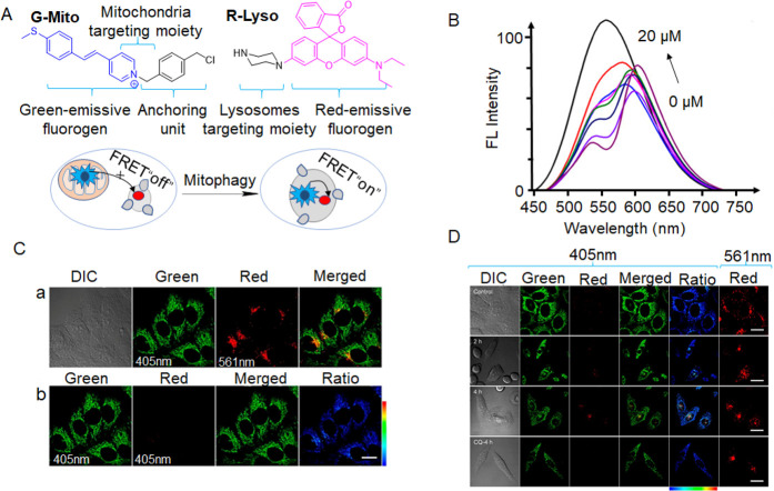
    1. She M.-T.; Yang J.-W.; Zheng B.-X.; Long W.; Huang X.-H.; Luo J.-R.; Chen Z.-X.; Liu A.-L.; Cai D.-P.; Wong W.-L.; et al. Design Mitochondria-Specific Fluorescent Turn-on Probes Targeting G-Quadruplexes for Live Cell Imaging and Mitophagy Monitoring Study. Chem. Eng. J. 2022, 446, 136947–136961. 10.1016/j.cej.2022.136947. - DOI
    1. Ding W. X.; Yin X. M. Mitophagy: Mechanisms, Pathophysiological Roles, and Analysis. Biol. Chem. 2012, 393, 547–564. 10.1515/hsz-2012-0119. - DOI - PMC - PubMed
    1. Li P.; Xiao H.; Cheng Y.; Zhang W.; Huang F.; Zhang W.; Wang H.; Tang B. A Near-Infrared-Emitting Fluorescent Probe for Monitoring Mitochondrial pH. Chem. Commun. 2014, 50, 7184–7187. 10.1039/c4cc01390e. - DOI - PubMed
    1. Wu M. Y.; Li K.; Liu Y. H.; Yu K. K.; Xie Y. M.; Zhou X. D.; Yu X. Q. Mitochondria-Targeted Ratiometric Fluorescent Probe for Real Time Monitoring of pH in Living Cells. Biomaterials 2015, 53, 669–678. 10.1016/j.biomaterials.2015.02.113. - DOI - PubMed

LinkOut - more resources