. 2024 Dec;11(48):e2407971.
doi: 10.1002/advs.202407971. Epub 2024 Oct 30.

LIF Promotes Sec15b-Mediated STAT3 Exosome Secretion to Maintain Stem Cell Pluripotency in Mouse Embryonic Development

Affiliations

LIF Promotes Sec15b-Mediated STAT3 Exosome Secretion to Maintain Stem Cell Pluripotency in Mouse Embryonic Development

Li Xu et al. Adv Sci (Weinh). 2024 Dec.

Abstract

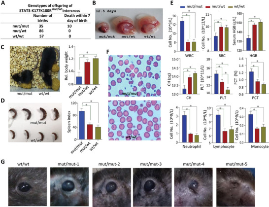
LIF maintains self-renewal growth in mouse embryonic stem cells (mESC) by activating STAT3, which translocates into nucleus for pluripotent gene induction. However, the ERK signaling pathway activated by LIF at large counteract with pluripotent gene induction during self-renewal growth. Here, it is reported that in mESC STAT3 undergoes multivesicular endosomes (MVEs) translocation and subsequent secretion, LIF-activated STAT3 is acetylated on K177/180 and phosphorylated on Y293 residues within the N-terminal coiled-coil domain, which is responsible for the interaction between STAT3 and Secl5b, an exocyst complex component 6B (EXOC6B). STAT3 translocation into MVEs resulted in the downregulation of T202/Y204-ERK1/2 phosphorylation and up-regulation of S9-GSK3β phosphorylation for maintaining mESC self-renewal growth. STAT3 with K177R/K180R or Y293F substitution fails to execute MVEs translocation and Secl5b-dependent secretion. Mice expressing K177RK180R substitution (STAT3mut/mut) are partially embryonic lethal. In STAT3mut/mut embryos, gene expressions related to hematological system function changed significantly and those living ones carry a series of abnormalities in the hematopoietic system. Furthermore, mice with Secl5b knockout exhibit embryonic lethality. Thus, Secl5b mediated STAT3 MVEs translocation regulates the balance of ERK and GSK3β signaling pathways and maintain mESC self-renewal growth, which is involved in regulating the stability of hematopoietic system.

Keywords: STAT3; Sec15b; cell pluripotency; exosome; multivesicular endosomes (MVEs).


Conflict of interest statement

The authors declare no conflict of interest.

Figures

Figure 1
Figure 1
STAT3 undergoes both nuclear and MVEs translocation in mouse embryos and mESC. A) Expression and distribution of STAT3 during preimplantation mouse development. STAT3 was stained with anti‐STAT3 antibody (red), and DNA was stained with 4,6‐diamidino‐2‐phenylindole (DAPI) (blue). B) Immune‐fluorescence staining of endogenous STAT3 in day‐4 and day‐9 outgrowths during mESC derivation. C) E14 cells maintained in 2i were treated with LIF (10 ng mL−1) for indicated time and separated for cytoplasmic and nuclear fractions. D) For the mESC maintained in LIF, STAT3, and DAPI were stained and visualized with confocal microscope. E) Immuno‐electron microscope demonstrated the distribution of STAT3. F) MVBs in the mESC were visualized with TEM. G) In 2i maintained E14 cells, MVBs were separated from whole cell lysates and analyzed by Western blotting.
Figure 2
Figure 2
Modifications in the CCD domain are key regulators for STAT3 MVEs translocation. A‐F) HEK293T cells were transiently transfected with STAT3‐GFP (A), STAT5a‐GFP (B), STAT3‐GFP fragments (C), GFP‐tagged STAT3/STAT5 domain swaps (D), GFP‐STAT3 with K177K180R or Y293F mutation (E) or GFP‐STAT3 with R31K or C108G mutation (F) and treated with or without LIF and stained with DAPI. G) The STAT3 protein crystal structure. The residues of R31, C108, K177, K180, and Y293 were indicated in different colors. H) HEK293T cells were transiently transfected with STAT3‐GFP followed by treatment with TSA (100 nM) for 6 h, then the cells were treated with or without LIF and stained with DAPI. I) GFP‐tagged STAT3‐R31K or STAT3‐C108G variant was co‐transfected with or without c‐Src in HEK293T cells. J) Myc‐STAT3 (1‐355) with or without Y293F substitution was co‐transfected with HA‐c‐Src in HEK‐293T cells. Purified STAT3 was analyzed in Western blot for tyrosine phosphorylation (pY20). K) Flag‐STAT3 (130‐585) with or without K177K180R double mutation was co‐transfected with CBP in HEK293T cells. Immunoprecipitated STAT3 was analyzed with pan acetyl‐lysine antibody in Western blot. L) STAT3 was immunoprecipited from CGR8 cells maintained in LIF or LIF withdrawal for 24 h and analyzed in Western blot with poly clonal antibodies prepared for STAT3‐K177 acetylation or ‐Y293 phosphorylation.
Figure 3
Figure 3
Sec15b facilitates STAT3 MVEs formation. A) Myc‐STAT3 was transiently transfected into cells, STAT3‐pull down proteins were purified by Myc Tag IP Kit and separated in 10% SDS‐PAGE, stained with coomassie brilliant blue followed by in‐gel trypsin digestion of targeted proteins and analysis of the resulting peptide fragments by mass spectrometry. B) Coimmunoprecipitation of STAT3 and Sec15b in the CGR8 mESC upon LIF withdrawal for indicated times. C) Myc‐tagged STAT3 full length (FL) or fragments were transfected with Flag‐Sec15b in HEK293T cells. Anti‐Flag immunoprecipitates were analyzed in Western blot for STAT3 and Sec15b interaction. D) The N‐terminal domains of both STAT3 and Sec15b were transfected into HEK293T cells, protein interaction was confirmed by Western blot. E) Structural analysis reveals a bundle of charged residues of the helical STAT3 N‐terminal domain is involved in the interaction with Sec15b N‐domain. F) STAT3‐Y293F, ‐K177K180R, ‐C108G, or ‐R31K mutants were cotransfected with Flag‐Sec15b in HEK293T cells, anti‐Myc immunoprecipitates were analyzed in Western blot for STAT3 and Sec15b interaction. G) Myc‐STAT3 and Flag‐Sec15b were transfected into HEK293T cells. Transport vesicles from the cytoplasm were isolated and were analyzed in Western blot for STAT3 and Sec15b MVEs translocation. H) The CGR8 mESC was treated with MNS at indicated concentrations for 2 h, protein interaction was checked in Western blot. I) Myc‐STAT3 and Flag‐Sec15b were cotransfected in HEK293T cells, then the cells were treated with BFA as indicated. STAT3 and Sec15b interaction was checked in Western blot. J) Immunostaining of STAT3 and Sec15b in the CGR8 mESC in the presence of LIF. K) Immunostaining of STAT3 and Sec15 in mESC D3 cells. L) Immunostaining of STAT3 and Sec15b in blastocyst on day 3.5.
Figure 4
Figure 4
LIF induced Sec15b‐STAT3 secretion. A) The CGR8 mESC was maintained in the presence or absence of LIF for indicated times. STAT3 was blotted from the secreted exosomes collected from the medium or from whole cell lysates. B) The relative amount of STAT3 secretion of (A) was quantitated. Data were shown as mean ± SD, n = 3. C) Diameter of the exosomes obtained from the mESC was determined by the nanosight system. Data were shown as mean ± SD, n = 3. D) Myc‐STAT3 was transiently transfected alone or together with Flag‐Sec15b in HEK293T cells followed by LIF treatment. Secreted exosomes collected from the medium were subjected to coimmunoprecipitation. Both the immunoprecipitates and whole cell lysates were checked in Western blot. E) HA‐Sec15a or HA‐Sec15b was transiently transfected in HEK293T cells followed by LIF treatment or no treatment. Secreted exosomes collected from the medium were subjected to Western blot analysis with STAT3 antibody. F) STAT3‐WT, ‐SY293F, ‐K177K180R, ‐C108G, or ‐R31K was transiently transfected in STAT3−/− MEFs followed by LIF treatment. Secreted exosomes and whole cell lysates were analyzed in Western blot. G) E14 mESC maintained in the LIF‐free medium (left) or LIF‐containing medium (middle and right) were visualized with TEM. In the absence of LIF, the niches between mESC were empty (left). In the presence of LIF, exosomes were visualized either in the niches between two cells (middle) or in the mESC ready for secretion into the niches (right). H) For the CGR8 mESC maintained in LIF, the exosome storm in secretion was visualized (left 1). The morphology of these isolated exosomes was visualized with TEM (left 2). Localization of gold beads with two different antibodies against STAT3 proteins in exosomes was visualized with immuno‐colloidal gold TEM (right 1 and 2). Localization of 10‐nm gold beads in exosomes was indicated (red arrow).
Figure 5
Figure 5
Sec15b‐STAT3 MVEs translocation and secretion maintain cellular pluripotency by modulating the ERK/GSK3β signaling pathway. A) Empty vector or STAT3 variants as indicated was overexpressed in STAT3−/− MEFs followed by LIF (20 ng mL−1) treatment for 30 min or not. GSK3α, GSK3β, and ERK phosphorylation were analyzed with indicated antibodies respectively in Western blot. B) The transcriptional activity of STAT3 variants was tested in dual SIE‐luciferase assay. Data were shown as mean ± SD, n = 3. C) Various STAT3 mutants were cotransfected with c‐Src into STAT3−/− MEF cells as indicated, and then western blot was performed by using different antibodies. D) Sec15b was knocked down with specific shRNA (shRNA‐453) in STAT3−/− mESC maintained with LIF. Both GSK3β (S9) and GSK3α (S21) phosphorylation were examined in Western blot. E) Quantitative RT‐PCR analysis of indicated gene expression in LIF‐maintained D3 cells after CTL shRNA or Secl5b specific shRNA was introduced for 72 h. The data were shown as mean + SD. F) The CGR8 mESC, pretreated in LIF withdrawal medium for 24 h, were treated with LIF or STAT3‐exosome (collected from D3 cells) for 30 min. PI3K‐pS473, AKT‐pT308, and GSK3β‐pS9 were estimated in Western blot with indicated antibodies. G) The CGR8 mESC, maintained in LIF‐free medium overnight, were treated with LIF or purified STAT3‐ exosomes for 72 h. RT‐PCR (left) and Western blot (right) were performed to examine indicated gene expression. H) The STAT3−/− mESC were treated with LIF + Smith 2i (3 µM CHIR99021 + 1 µM PD0325901), LIF + CHIR99021, LIF + PD0325901, LIF alone, STAT3 exosome alone (5 mg mL−1), or LIF + STAT3‐Sec15 exosome for 48 h. The ERK phosphorylation was analyzed in Western blot. I) The STAT3−/− mESC colonial growth under different conditions: LIF, LIF + Smith 2i, LIF + CHIR99021, LIF + PD0325901, LIF + STAT3‐exosome (5 mg mL−1) and STAT3‐exosome. J) Exosomes were isolated from CGR8 mESCs and STAT3−/− mESCs. The expression levels of STAT3, Lamin B1, and TSG101 in exosome and whole cell lysate (WCL) were analyzed by Western blot using the specified antibodies. K) The CGR8 mESC colonial growth under different conditions: LIF withdrawal, LIF (1000 U mL−1), STAT3‐exosome (5 mg mL−1) obtained from D3 mESC, and exosome (5 mg mL−1) obtained from STAT3−/− mESC (upper panel).
Figure 6
Figure 6
Mice with STAT3‐K177K180R mutant exhibit significant developmental abnormalities. A) The statistical results of offspring genotyping of STAT3‐K177K180R intercross. B) Abnormal phenotypes of STAT3‐K177K180R homozygous (STAT3 mut/mut ) embryo on 12.5 days. C) The STAT3 mut/mut mice exhibit slow growth, with both reduced weight and smaller size (letf). The body weight (n = 12) of STAT3 wild type (STAT3 wt/wt ), heterozygous (STAT3 mut/wt ), or homozygous (STAT3 mut/mut ) mice in 12 weeks (right). D) The STAT3 mut/mut mice display splenomegaly (left). The spleen index (n = 6) of STAT3 wild type (STAT3 wt/wt ), heterozygous (STAT3 mut/wt ), or homozygous (STAT3 mut/mut ) mice in 12 weeks (right). E) The number of different categories of peripheral blood cells from STAT3 mut/mut , STAT3 mut/wt , and STAT3 wt/wt mice were analyzed with an automated hematology analyzer (n = 11). F) Red blood cell staining of STAT3 wt/wt and STAT3 mut/mut mice. G) The STAT3 mut/mut mice develop eye abnormalities and even lead to atrophy and blindness in later growth stages.
Figure 7
Figure 7
STAT3‐K177K180R mutation in mice exhibit reduced embryonic stem cell pluripotency and hematopoietic abnormalities. A) Analysis of differentially expressed genes regulating pluripotency in STAT3 mut/mut mouse embryos. Downregulated genes are shown in green, and upregulated genes are displayed in red. B) Heat map of related gene expression. Most of genes involved in regulating pluripotency of mouse stem cells and mesoderm development were down‐regulated in STAT3 mut/mut embryos. In contrast, some genes involved in MAPK pathway and ectoderm development were up‐regulated. C) Ingenuity Pathway Analysis (IPA)‐identified top 3 most significant gene networks with score ≥30. D) IPA network analysis of differentially expressed genes related hematological system development. E) In vivo differentiation of Th17 cells and Treg cells from STAT3 mut/mut , STAT3 mut/wt , and STAT3 wt/wt mice were detected with FACS. F) The statistical results of offspring genotypes from Sec15b+/− intercross.

References

    1. Evans M. J., Kaufman M. H., Nature 1981, 292, 154. - PubMed
    1. Smith A. G., Heath J. K., Donaldson D. D., Wong G. G., Moreau J., Stahl M., Rogers D., Nature 1988, 336, 688. - PubMed
    1. Niwa H., Burdon T., Chambers I., Smith A., Genes Dev. 1998, 12, 2048. - PMC - PubMed
    1. Raz R., Lee C. K., Cannizzaro L. A., d'Eustachio P., Levy D. E., Proc Natl Acad Sci U S A 1999, 96, 2846. - PMC - PubMed
    1. Niwa H., Ogawa K., Shimosato D., Adachi K., Nature 2009, 460, 118. - PubMed

MeSH terms

LinkOut - more resources