. 2024 Nov 5;121(45):e2404947121.
doi: 10.1073/pnas.2404947121. Epub 2024 Oct 30.

Hyponatremia unleashes neutrophil extracellular traps elevating life-threatening pulmonary embolism risk

Affiliations

Hyponatremia unleashes neutrophil extracellular traps elevating life-threatening pulmonary embolism risk

Minoru Inoue et al. Proc Natl Acad Sci U S A. .

Abstract

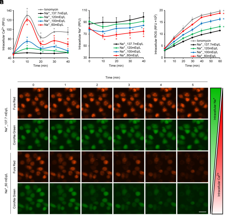
Neutrophil extracellular traps (NETs), essential for controlling infections, can induce various pathologies when dysregulated. Known triggers for infection-independent NETs release exist, yet a comprehensive understanding of the conditions prompting such responses is lacking. In this study, we identify hyponatremia as an independent inducer of NETs release, a common clinical condition that disrupts sodium/calcium exchange within neutrophils. This disruption leads to an excess of intracellular calcium, subsequent elevation of reactive oxygen species (ROS), and the citrullination of histone H3, culminating in the activation of NETs-release pathways. Notably, under hyponatremic conditions, this mechanism is exacerbated during infectious states, leading to the deposition of NETs in the lungs and increasing the risk of life-threatening pulmonary embolism. Our findings underscore the critical role of sodium and calcium homeostasis in neutrophil functionality and provide insights into the pathogenesis of hyponatremia-associated diseases, highlighting potential therapeutic interventions targeting NETs dynamics.

Keywords: SARS-CoV-2; hyponatremia; neutrophil extracellular traps; pulmonary embolism; sodium–calcium exchanger.


Conflict of interest statement

Competing interests statement:The authors declare no competing interest.

Figures

Fig. 1.
Fig. 1.
Hyponatremic condition inhibits forward-mode Na+/Ca2+ exchange and elevates ROS levels within neutrophils. (AD) Human neutrophils were cultured in media with modified Na+ concentrations as indicated. A normonatremic condition (i.e., 137.7 mEq/L) with 5 μM Ionomycin treatment was used as a positive control for the induction of Ca2+ influx. (A and B) Changes in intracellular Ca2+ and Na+ levels, measured using the fluorescent indicators Fluo-4 AM for Ca2+ (A) and CoroNa Green AM for Na+. (C) Neutrophils were labeled with indicators for Ca2+ (Fura Red AM) and Na+ (CoroNa Green AM) under normonatremic culture conditions. Representative images are shown of neutrophils cultured in normonatremic (i.e., 137.7 mEq/L) and hyponatremic (i.e., 60 mEq/L) culture media. Notably, Fura Red AM exhibits a decrease in fluorescence intensity upon an increase in intracellular Ca2+ levels when excited at a wavelength of 531 nm, a behavior contrasting with that of Fluo-4 AM. Bar = 20 μm. (D) Changes in intracellular ROS levels represented by the fluorescent intensity of dichlorodihydrofluorescein diacetate (DCFDA). (A, B, and D) Results represent the mean ± SD (n = 3; biological replicates, significant differences compared with the normonatremic control at *P <0.05 by Dunnett’s test).
Fig. 2.
Fig. 2.
Na+ deficiency promotes NETs release via ROS-mediated peptidyl arginine deiminase (PAD) activation. (AF) Human neutrophils were incubated in Na+-adjusted medium for 5 h. A normonatremic condition (i.e., 137.7 mEq/L) with 75 μM EIPA treatment served as a positive control for NETs induction via Na+/Ca2+ exchange inhibition. To counteract intracellular ROS or inhibit PAD activation, neutrophils were pretreated with 50 μM glutathione ethyl ester (GSHee) or 10 μM BB-Cl-amidine (BB-CLA) 10 min before exposure to severe hyponatremia (i.e., 60 mEq/L). (A) Representative images of neutrophils stained with the cell-permeable DNA dye Hoechst 33342 (blue) and cell-impermeable DNA dye SytoxOrange (red). Bar = 100 μm. (B and E) Representative immunofluorescence images stained with DAPI (blue) and anti-citrullinated histone H3 (Cit-H3, green). Bar = 50 μm. (C, D and F) NETs levels in cell culture supernatants from neutrophils under the specified culture conditions. (C, D and F) Data represent mean ± s.d. (n = 3; biological replicates; significant differences compared to the normonatremic control at *P < 0.05 by Dunnett’s test (C); and between the two indicated groups at *P <0.05 by Student’s t-test (D and F).
Fig. 3.
Fig. 3.
Enhanced SARS-CoV-2-driven NETs release under mild hyponatremic conditions. Human neutrophils were cultured in Na+-adjusted media, with or without SARS-CoV-2, for 5 h (A) Representative immunofluorescence images depict neutrophils stained with DAPI (blue) and anti-citrullinated histone H3 (Cit-H3, green). A normonatremic condition (i.e., 137.7 mEq/L) with 5 μM Ionomycin treatment was used as a positive control. Bar = 25 μm. (B) NETs levels were quantified in supernatants from neutrophils under the specified conditions. Data are presented as mean ± s.d. (n = 3; biological replicates; significant differences compared with the three indicated groups at *P <0.05 by Dunnett’s test).
Fig. 4.
Fig. 4.
Lung-specific deposition of NETs induced by hyponatremia. C57BL/6 N mice received intraperitoneal injection of sterile distilled water (0.15 mL/g body weight) and a subcutaneous dose of 1 mg (deamino-Cys1, D-Arg8)-Vasopressin acetate salt hydrate (dDAVP) in 50 μL saline. Control mice were administered an equivalent volume of saline intraperitoneally as a mock treatment. (A) Plasma Na+ concentration variations post-injection. (B) NETs quantification in plasma collected 12 h following hyponatremia induction. (C) A phosphate-buffered saline (PBS) solution of SytoxGreen (5 μL in 200 μL PBS) was injected into the tail vein 12 h after hyponatremia induction. The brain, lungs, and heart were harvested post-SytoxGreen injection and immediately imaged using two-photon excitation microscopy. SytoxGreen-positive elongated structures are indicative of NETs. Bar = 40 μm. Data are expressed as mean ± s.d. (n = 3, significant differences compared with the uninjected control at *P <0.05 by Dunnett’s test)
Fig. 5.
Fig. 5.
Heightened risk of fatal pulmonary thrombosis by hyponatremia during sepsis induced by lipopolysaccharide. C57BL/6 N mice and green fluorescent protein (GFP)-transgenic mice were subjected to intraperitoneal injection of LPS (17.5 μg/g body weight) alongside sterile distilled water (0.15 mL/g body weight) and a subcutaneous injection of 1 mg (deamino-Cys1, D-Arg8)-Vasopressin acetate salt hydrate (dDAVP) in 50 μL saline. Saline-injected mice (0.15 mL/g body weight intraperitoneally) served as mock-treated controls. Where indicated, DNase I (7.5 KU in 100 μL PBS) was administered at 2 and 6 h post-induction, or thrombin (50 U in 100 μL PBS) was injected via the tail vein. (A) Masson’s trichrome staining of lung tissue from C57BL/6 N mice harvested 12 h post-induction. The lower panel shows a magnified view of the area outlined by the dotted square in the upper panel. Bar = 100 μm. (B) Serum D-dimer levels at 12 h post-induction in C57BL/6 N mice (n = 6 per group). *P <0.05 by Dunnett’s test. (C) 12 h following the induction of hyponatremia and/or sepsis, a PBS solution with SytoxOrange (5 μL in 200 μL PBS) was administered via the tail vein in GFP-transgenic mice. Intravital lung imaging was conducted using a two-photon excitation microscope. Representative lung images are displayed, NETs-like SytoxOrange-positive structures in magenta. Bar = 50 μm. (D) Mouse survival following the concurrent induction of hyponatremia and sepsis in C57BL/6 N mice (n = 12 per group) was analyzed by Kaplan–Meier log-rank survival analysis. *P <0.05 (LPS vs. DDW+dDAVP+LPS).

References

    1. Kolaczkowska E., Kubes P., Neutrophil recruitment and function in health and inflammation. Nat. Rev. Immunol. 13, 159–175 (2013). - PubMed
    1. Brinkmann V., et al. , Neutrophil extracellular traps kill bacteria. Science 303, 1532–1535 (2004). - PubMed
    1. Lood C., et al. , Neutrophil extracellular traps enriched in oxidized mitochondrial DNA are interferogenic and contribute to lupus-like disease. Nat. Med. 22, 146–153 (2016). - PMC - PubMed
    1. Pilsczek F. H., et al. , A novel mechanism of rapid nuclear neutrophil extracellular trap formation in response to Staphylococcus aureus. J. Immunol. 185, 7413–7425 (2010). - PubMed
    1. Yipp B. G., et al. , Infection-induced NETosis is a dynamic process involving neutrophil multitasking in vivo. Nat. Med. 18, 1386–1393 (2012). - PMC - PubMed

LinkOut - more resources