

Melatonin associated with bacterial cellulose-based hydrogel improves healing of skin wounds in diabetic rats
- PMID: 39476070
- PMCID: PMC11506694
- DOI: 10.1590/acb399024
Melatonin associated with bacterial cellulose-based hydrogel improves healing of skin wounds in diabetic rats
Abstract
Purpose: To describe the effects of melatonin associated with bacterial cellulose-based hydrogel on healing of skin wounds in diabetic rats.
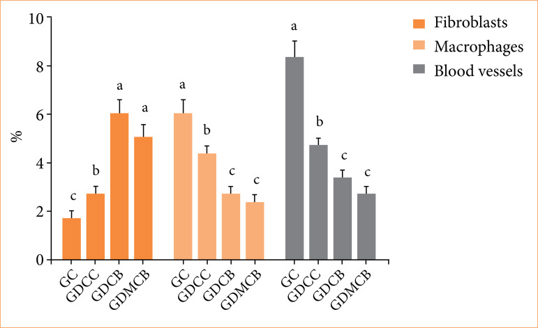
Methods: Streptozotocin was used to induce diabetes in Wistar rats. After wound induction, animals were randomly divided into groups GC, GDCC, GDCB, and GDMCB. Animals were evaluated in days 3, 7, and 14 for the following variables: glycemic levels, histopathological and histochemical analyses, healing rate, morphometry and C-reactive protein.
Results: There was no change in glycemic levels in the diabetic animals as a result of the treatments; histopathological analyses showed better healing in GDCB and GDMCB groups, as well as histochemistry; at day 14, the highest healing rate was observed in animals from the GDMCB group, reaching almost 100%; morphometry revealed a significant increase of fibroblasts and reduction of macrophages and blood vessels in lesions treated with bacterial cellulose associated or not with melatonin when compared to the other experimental groups. There was also an increase in C-reactive protein in GDCC group at day 14.
Conclusion: Bacterial cellulose-based dressings associated with systemic melatonin showed beneficial results in experimentally induced wounds in diabetic rats, favoring the healing process.
Conflict of interest statement
Figures


Similar articles
-
In-vivo evaluation of Alginate-Pectin hydrogel film loaded with Simvastatin for diabetic wound healing in Streptozotocin-induced diabetic rats.Int J Biol Macromol. 2021 Feb 28;171:308-319. doi: 10.1016/j.ijbiomac.2020.12.221. Epub 2021 Jan 7. Int J Biol Macromol. 2021. PMID: 33421467
-
Treatment for diabetic ulcer wounds using a fern tannin optimized hydrogel formulation with antibacterial and antioxidative properties.J Ethnopharmacol. 2016 Aug 2;189:277-89. doi: 10.1016/j.jep.2016.05.032. Epub 2016 May 18. J Ethnopharmacol. 2016. PMID: 27208868
-
[Preliminary evaluation and mechanism of adipose-derived stem cell transplantation from allogenic diabetic rats in the treatment of diabetic rat wounds].Zhonghua Shao Shang Za Zhi. 2019 Sep 20;35(9):645-654. doi: 10.3760/cma.j.issn.1009-2587.2019.09.002. Zhonghua Shao Shang Za Zhi. 2019. PMID: 31594182 Chinese.
-
Topical application of melatonin accelerates the maturation of skin wounds and increases collagen deposition in a rat model of diabetes.J Tissue Viability. 2022 Nov;31(4):606-613. doi: 10.1016/j.jtv.2022.07.015. Epub 2022 Aug 23. J Tissue Viability. 2022. PMID: 36068126
-
Bacterial cellulose membrane associated with red propolis as phytomodulator: Improved healing effects in experimental models of diabetes mellitus.Biomed Pharmacother. 2019 Apr;112:108640. doi: 10.1016/j.biopha.2019.108640. Epub 2019 Feb 20. Biomed Pharmacother. 2019. PMID: 30784929
Cited by
-
Exo-hydrogel therapy: a revolutionary approach to managing diabetic complications.J Nanobiotechnology. 2025 Aug 11;23(1):558. doi: 10.1186/s12951-025-03621-6. J Nanobiotechnology. 2025. PMID: 40790200 Free PMC article. Review.
References
MeSH terms
Substances
LinkOut - more resources
Full Text Sources
Research Materials
Miscellaneous
