. 2024 Oct 30;40(1):93.
doi: 10.1007/s10565-024-09931-z.

NPR1 promotes cisplatin resistance by inhibiting PARL-mediated mitophagy-dependent ferroptosis in gastric cancer

Affiliations

NPR1 promotes cisplatin resistance by inhibiting PARL-mediated mitophagy-dependent ferroptosis in gastric cancer

Chengwei Wu et al. Cell Biol Toxicol. .

Abstract

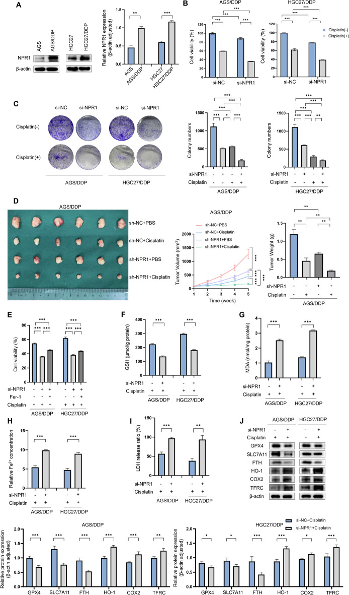
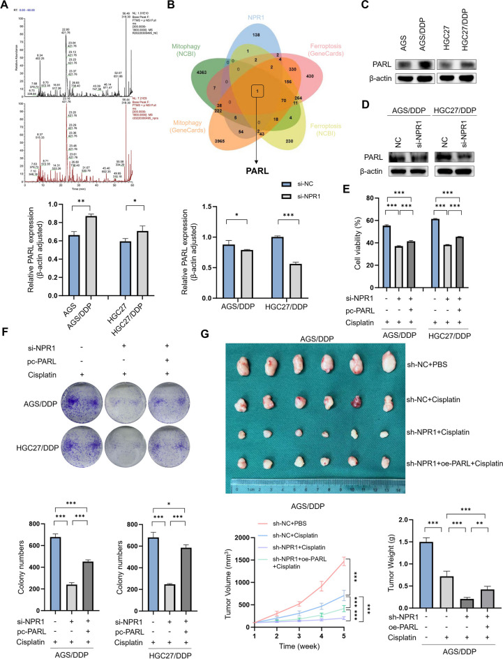
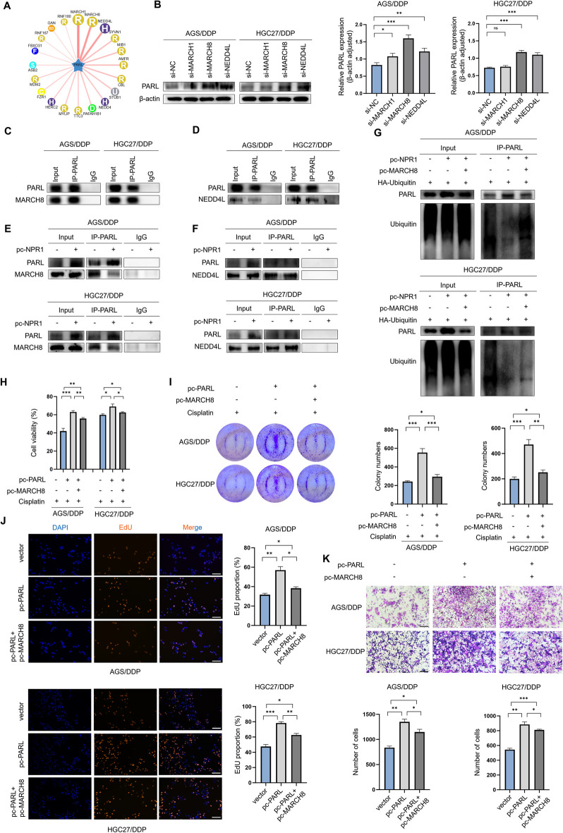
Cisplatin-based chemotherapy serves as the standard of care for individuals with advanced stages of gastric cancer. Nevertheless, the emergence of chemoresistance in GC has detrimental impacts on prognosis, yet the underlying mechanisms governing this phenomenon remain elusive. Level of mitophagy and ferroptosis of GC cells were detected by fluorescence, flow cytometry, GSH, MDA, Fe2+ assays, and to explore the specific molecular mechanisms between NPR1 and cisplatin resistance by performing western blot and coimmunoprecipitation (co-IP) assays. These results indicates that NPR1 positively correlated with cisplatin-resistance and played a crucial part in conferring resistance to cisplatin in gastric cancer cells. Mechanistically, NPR1 affected levels of mitophagy and ferroptosis in human cisplatin-resistance GC cells with cisplatin treatment. Specifically, NPR1 inhibited mitophagy-dependent ferroptosis by reducing the ubiquitination-mediated degradation of PARL; moreover, NPR1 promoted PARL stabilization by disrupting the PARL-MARCH8 complex, which ultimately led to the development of chemoresistance in GC cells. Considering our findings, NPR1 appears to play an important role in chemotherapy for GC. NPR1 could potentially be used to overcome chemotherapy resistance.

Keywords: Chemoresistance; Ferroptosis; Gastric Cancer; Mitophagy; NPR1; PARL; Ubiquitination.


Conflict of interest statement

The authors declare no competing interests.

Figures

Fig. 1
Fig. 1
NPR1 affects Cisplatin resistance in gastric cancer in vivo and vitro through a pathway that influences ferroptosis. A Differential expression of NPR1 in cisplatin-resistant and sensitive cell lines of gastric cancer detected by western blot assay. B-C CCK8 assay detects cisplatin resistance in AGS/DDP and HGC27/DDP cell lines after knockdown of NPR1. Colony formation assay detects cisplatin resistance in AGS/DDP and HGC27/DDP cells lines after knockdown of NPR1.  Cisplatin (6 µM). D Mouse subcutaneous tumor formation assay detects cisplatin resistance in AGS/DDP after knockdown of NPR1. Cisplatin (5 mg/kg, three times a week). E CCK8 assay detects cisplatin resistance in AGS/DDP and HGC27/DDP cell lines after knockdown of NPR1 and concomitant addition of Fer-1. F-I Analysis of glutathione (GSH), malondialdehyde (MDA), Fe2+, and lactate Dehydrogenase (LDH) levels detect ferroptosis level in AGS/DDP and HGC27/DDP cell lines with knockdown of NPR1. J Expression of ferroptosis-related proteins after knockdown of NPR1 in AGS/DDP and HGC27/DDP cell lines by western blot assay
Fig. 2
Fig. 2
NPR1 induced ferroptosis occurs in gastric cancer cells via a mitophagy-dependent pathway. A Immunofluorescence assay detect the fluorescent signal intensity of LC3B and the co-localisation of LC3B with Mitotrack after knockdown of NPR1 in AGS/DDP and HGC27/DDP (scale bar: 5 μm). B Immunofluorescence assay detect the co-localisation of mitochondria and lysosomes after knockdown of NPR1 (scale bar: 5 μm). C JC-1 assay after knockdown of NPR1 (scale bar: 20 μm). D Western blot assay detect the levels of mitophagy-related proteins and ferroptosis-related proteins after treatment with si-NPR1. E Flow cytometric assay detects ROS levels due to knockdown of NPR1 (MFI: Mean Fluorescence Intensity). F Western blot assay detect the expression levels of mitophagy-related proteins after treatment with autophagy inhibitor 3-MA. G The expression of Drp1 and MFN1 after knockdown of si-NPR1 in AGS/DDP, HGC27/DDP and AGS, HGC27 cells. H-J Analysis of MDA, GSH, and Fe2+ levels showed that the elevated levels of ferroptosis due to knockdown of NPR1. K. Western blot assay detect the expression levels of ferroptosis-related proteins after treatment with mitophagy inhibitor Mdivi-1
Fig. 3
Fig. 3
NPR1 promotes Cisplatin resistance through PARL. A-B By overexpressing NPR1 in the GC cell line AGS for IP/MS, which yielded proteins interacting with NPR1 with pull-downs, and by intersecting with the mitophagy and ferroptosis databases of NCBI (https://www.ncbi.nlm.nih.gov/gene) and GENECARDS (https://www.genecards.org/). C Differential expression of PARL in cisplatin-resistant and sensitive cell lines of gastric cancer detected by western blot assay. D Western blot assay showed that compared with the control groups that knockdown of NPR1 in AGS/DDP and HGC27/DDP cells significantly reduced PARL expression. E CCK8 assay showed that knockdown of NPR1 inhibited cisplatin resistance in AGS/DDP and HGC27/DDP, and overexpression of PARL reversed the decrease in cisplatin resistance caused by knockdown of NPR1. F Colony formation assays showed that PARL reverted the reduced cisplatin resistance of AGS/DDP and HGC27/DDP due to knockdown of NPR1, and overexpression of PARL reversed the decrease in cisplatin resistance caused by knockdown of NPR1. G In subcutaneous tumor formation experiments in mice, the decrease in tumor size, volume, and weight due to knockdown of NPR1 are all reverted by overexpression of PARL
Fig. 4
Fig. 4
NPR1 promotes mitophagy-dependent ferroptosis through PARL. A Immunofluorescence assay detect the fluorescent signal intensity of LC3B and the co-localisation of LC3B with Mitotrack after knockdown of NPR1 and overexpression of PARL in AGS/DDP and HGC27/DDP (scale bar: 5 μm). B Immunofluorescence assay detect the co-localisation of mitochondria and lysosomes after knockdown of NPR1 and overexpression of PARL (scale bar: 5 μm). C JC-1 assay after knockdown of NPR1 and overexpression of PARL (scale bar: 20 μm). D Western blot assays detect the levels of mitophagy-related proteins after knockdown of NPR1 and overexpression of PARL. E Flow cytometric assay detects ROS levels after knockdown of NPR1 and overexpression of PARL. F–H Analysis of MDA, GSH, and Fe2+ levels showed that the levels of ferroptosis after knockdown of NPR1 and overexpression of PARL. I Western blot assays detect the levels of ferroptosis-related proteins after knockdown of NPR1 and overexpression of PARL
Fig. 5
Fig. 5
NPR1 affects the ubiquitination process of PARL. A RT-qPCR detects PARL mRNA levels after knockdown of NPR1. B-C Immunofluorescence co-localisation assay and co-IP detect the existence of interaction and co-localisation between NPR1 and PARL in GC cells (scale bar: 5 μm). D Western blot assays examine the half-life of PARL in GC cells treated with the protein synthesis inhibitor cycloheximide (CHX, 200 µg/mL). E Western blot assays detect effect on NPR1-regulated PARL expression by autophagy inhibitor chloroquine (20 µM) and 26S protostome inhibitor MG-132 (10 µM). F co-IP assay detects the ubiquitin signals of PARL that after endogenous PARL was immunoprecipitated in cells transfected with HA-Ub in NPR1-siRNA cells
Fig. 6
Fig. 6
NPR1 affects the process of ubiquitination of PARL by competing with MARCH8 for the binding of PARL. A UbiBrowser (http://ubibrowser.bio-it.cn/ubibrowser) analyze the E3 ligase that interacts with PARL using a computational predictive system. B Western blot analysis revealed the impact on PARL expression after knocking down MARCH1, MARCH8, and NEDD4L in AGS/DDP and HGC27/DDP cells, compared with the control group. C-D co-IP assay confirmed the existence of interaction between MARCH8 and PARL, NEDD4L and PARL in GC cells. E–F co-IP detected E3 ligase that competitively binds to PARL with NPR1, less MARCH8 protein was precipitated with PARL in the overexpression of NPR1 cells compared with the control GC cells. G co-IP assay detects the ubiquitin signals of PARL that after endogenous PARL was immunoprecipitated in cells transfected with HA-Ub in pc-NPR1 and pc-MARCH8. H-I The CCK8 assay and colony formation assay indicates the functions in drug resistance of PARL and its E3 ubiquitin ligase MARCH8. J-K The EdU assay and transwell assay indicates the functions in tumorigenesis of PARL and its E3 ubiquitin ligase MARCH8 (scale bar: 100 μm)

References

    1. Ajani JA, et al. Gastric Cancer, Version 2.2022, NCCN clinical practice guidelines in oncology. J Natl Compr Canc Netw. 2022;20:167–92. 10.6004/jnccn.2022.0008. - PubMed
    1. Cao T, et al. NPRA promotes fatty acid metabolism and proliferation of gastric cancer cells by binding to PPARα. Transl Oncol. 2023;35:101734. 10.1016/j.tranon.2023.101734. - PMC - PubMed
    1. Chen X, Kang R, Kroemer G, Tang D. Broadening horizons: the role of ferroptosis in cancer. Nat Rev Clin Oncol. 2021;18:280–96. 10.1038/s41571-020-00462-0. - PubMed
    1. Chen Z, et al. MSC-NPRA loop drives fatty acid oxidation to promote stemness and chemoresistance of gastric cancer. Cancer Lett. 2023;565:216235. 10.1016/j.canlet.2023.216235. - PubMed
    1. Dai E, et al. Autophagy-dependent ferroptosis drives tumor-associated macrophage polarization via release and uptake of oncogenic KRAS protein. Autophagy. 2020;16:2069–83. 10.1080/15548627.2020.1714209. - PMC - PubMed

Publication types

MeSH terms

LinkOut - more resources