. 2024 Oct 31;15(1):9406.
doi: 10.1038/s41467-024-53730-3.

Generation of binder-format-payload conjugate-matrices by antibody chain-exchange

Affiliations

Generation of binder-format-payload conjugate-matrices by antibody chain-exchange

Vedran Vasic et al. Nat Commun. .

Abstract

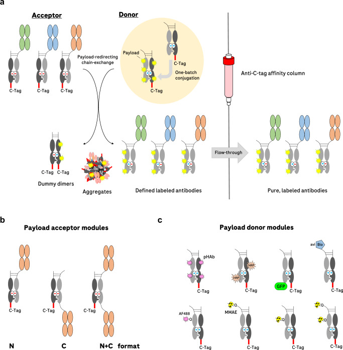
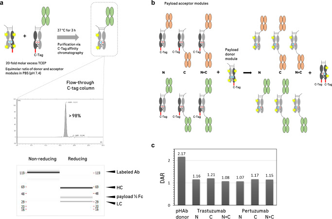
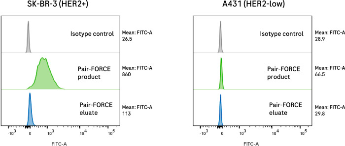
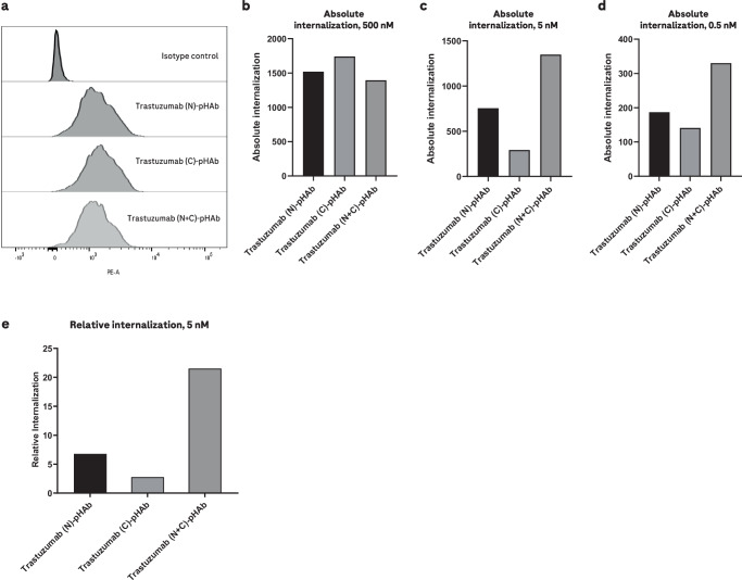
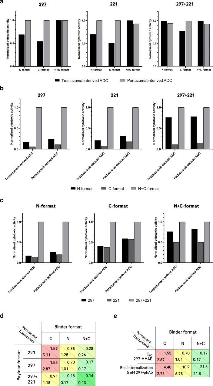
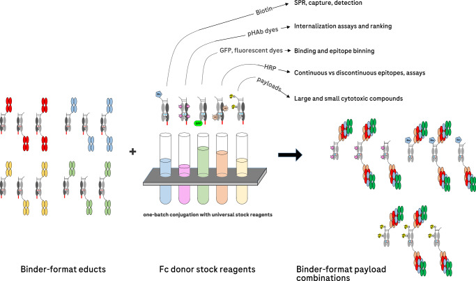
The generation of antibody-drug conjugates with optimal functionality depends on many parameters. These include binder epitope, antibody format, linker composition, conjugation site(s), drug-to-antibody ratio, and conjugation method. The production of matrices that cover all possible parameters is a major challenge in identifying optimal antibody-drug conjugates. To address this bottleneck, we adapted our Format Chain Exchange technology (FORCE), originally established for bispecific antibodies, toward the generation of binder-format-payload matrices (pair-FORCE). Antibody derivatives with exchange-enabled Fc-heterodimers are combined with payload-conjugated Fc donors, and subsequent chain-exchange transfers payloads to antibody derivatives in different formats. The resulting binder-format-conjugate matrices can be generated with cytotoxic payloads, dyes, haptens, and large molecules, resulting in versatile tools for ADC screening campaigns. We show the relevance of pair-FORCE for identifying optimal HER2-targeting antibody-drug conjugates. Analysis of this matrix reveals that the notion of format-defines-function applies not only to bispecific antibodies, but also to antibody-drug conjugates.


Conflict of interest statement

All coauthors are either employed by Roche or were previously employed by Roche at the time the work was conducted. Roche has an interest in, and patents/patent applications including V.V., S. Dickopf, K.M., H.D., S. Dengl, and U.B. as inventors that relate to FORCE technologies and antibody engineering.

Figures

Fig. 1
Fig. 1. Overview of the payload-redirecting chain-exchange concept (pair-FORCE).
a Different binders (in green, blue, and orange) have varying sequences in their Fab regions. On the upper right (highlighted in the yellow circle), a single conjugation reaction with an Fc donor molecule is depicted. The conjugated donor molecule is then applied to the exchange reaction with each of the different binders (acceptors) in the presence of the reducing agent TCEP, which reduces the disulfide bridges in the hinge region (shown as dashed lines). After the chain-exchange reaction, the mixture is applied to a C-tag affinity column, which captures dummy-dimers, aggregates, and non-reacted educt molecules. The products - labeled antibodies with a defined composition —are present in the flow-through with high purity. The hinge disulfides of the product antibodies reoxidize after C-tag purification. b Antibody-derived acceptor molecules can be produced in three different formats (N, C, and N + C) and can be combined with different payload-donor entities. c A variety of payload-donor modules can be produced through nonspecific conjugation or site-specific conjugation approaches (depicted here as either biotinylation of an Avi tag by the BirA enzyme or transglutaminase-mediated Q-tag conjugation). After pairing with binder-containing acceptor modules through pair-FORCE, the labeled antibody derivatives can be utilized for a variety of approaches, including monitoring of internalization, immunoblotting, FACS, epitope binning, SPR assays, immunoprecipitation, and ADC cytotoxicity assays. pHAb pH-sensitive dye, HRP horseradish peroxidase, GFP green fluorescent protein, avi recognition sequence for site-specific biotinylation, AF488 Alexa Fluor 488, MMAE monomethyl auristatin E.
Fig. 2
Fig. 2. Chain-exchange-mediated attachment of fluorescent dyes from NHS-conjugated donor modules with pair-FORCE.
a Upper panel: reactant molecules are mixed in equimolar concentrations in PBS under mild reducing conditions (20-fold molar excess TCEP). The mixture is incubated at 37 °C for 3 h while shaking. The reaction is then loaded onto a C-tag affinity column and the product is collected in the flow-through. Lower panel: The labeled product, a HER2 binder (Trastuzumab-derived) in the N-format with conjugated pHAb fluorescent dye molecules, has a purity of >98% as measured by analytical SEC. The capillary electrophoresis (CE-SDS) reveals defined bands of expected sizes under both non-reducing and reducing conditions. HC heavy chain, LC light chain, payload ½ Fc payload-conjugated Fc chain, Ab antibody. b A binder-format matrix of two different HER2 binders (derived from Trastuzumab and Pertuzumab) in three different formats (N, C, and N + C) was combined in a pair-FORCE reaction with an Fc donor molecule that was conjugated with pHAb (a pH-sensitive fluorophore) through NHS-amine labeling. c Analysis of the pair-FORCE products via mass spectrometry reveals a similar DAR (drug-to-antibody ratio) for all products, irrespective of the format. As expected, the pHAb-conjugated Fc donor molecule (Payload-donor module in 1c) has an approximately twofold greater DAR than the product molecules. Shown are results from one independent experiment (n = 1), see Methods section for details.
Fig. 3
Fig. 3. Antibody-GFP fusion proteins can be generated by pair-FORCE.
EGFP can be transferred to antibody derivatives in different formats by pair-FORCE and can be applied to assess binding and the presence of target antigens by flow cytometry (see Methods section for flow cytometry details). A C-terminal EGFP molecule was transferred via pair-FORCE from a donor Fc-EGFP fusion to a HER2 binder-acceptor module in the N-format (derived from Trastuzumab). The pair-FORCE product (C-tag column flow-through) and pair-FORCE eluate (C-tag column eluate, contains by-products and unreacted Fc donor molecules) were incubated with HER2-positive SK-BR-3 cells or HER2-low A431 cells, and binding was determined by flow cytometry. EGFP fluorescence was monitored in the FITC channel. The successful transfer of EGFP to the Trastuzumab acceptor module was confirmed by the increased EGFP fluorescence on HER2-positive SK-BR-3 cells treated with the pair-FORCE product. Target-specific binding was retained after pair-FORCE, as only minimal fluorescence was detected on HER2-low A431 cells that were treated with the pair-FORCE product (right panel). FITC fluorescein isothiocyanate.
Fig. 4
Fig. 4. Internalization of pair-FORCE-generated HER2 antibodies in different formats on HER2-expressing SK-BR-3 cells.
Internalization of Trastuzumab-derived HER2 binders in the N, C, and N + C format by HER2-expressing SK-BR-3 cells was assessed by flow cytometry using HER2 antibodies conjugated with the pH-sensitive dye pHAb. The conjugated HER2 antibodies were generated with pair-FORCE technology. The fluorescence of the pHAb dye is poor at neutral pH but increases substantially upon trafficking into the acidic environment of the endo-/lysosomal pathway. For all panels, n = 1 independent replicates were performed. a Fluorescence histogram from flow cytometry experiments of SK-BR-3 cells treated with pHAb-conjugated HER2 binders at a saturating concentration of 500 nM. pHAb fluorescence was measured in the PE (phycoerythrin) channel. b The same experiment as (a), but absolute internalization is depicted as a bar graph. Absolute internalization is defined as the geometric mean of pHAb fluorescence in flow cytometry experiments. c Absolute internalization of pHAb-conjugated HER2 antibodies as in (b), but at a concentration of 5 nM. d Absolute internalization of pHAb-conjugated HER2 antibodies as in (b, c), but at a concentration of 0.5 nM. e Relative internalization of Trastuzumab-derived HER2 antibodies at a concentration of 5 nM. Relative internalization is calculated as the ratio of absolute internalization to absolute binding, which is derived from flow cytometry binding experiments with AF488-labeled HER2 antibodies generated by pair-FORCE (see Methods section for details). The absolute binding is defined as the geometric mean of AF488 fluorescence of SK-BR-3 cells treated with 200 nM of AF488-conjugated HER2 antibodies. N, C, and N + C refer to the binder formats in Fig. 1b. Source data for Fig. 4b–e are provided as a Source Data file.
Fig. 5
Fig. 5. Influence of individual and combined parameters on HER2-MMAE ADC activity.
In Fig. 5a–c, in order to define rules for each variable, the highest activity (i.e., the lowest IC50 value in cell proliferation/cytotoxicity assays) for a set of variables in each individual subgroup was set to 1.0, and the respective activities (IC50 values) were normalized relative to this value. This allows for the determination of the relative influence of the HER2 binder, format, and conjugation position/DAR on ADC activity. Relative values are comparable only within the same subgroup in each bar graph. Parameters that are compared in each panel are listed in the legend below the bar graphs. The values 297 and 221 refer to the MMAE conjugation site (Q-tag at positions 297 and 221 in the Fc donor productive chain). Individual data sets underlying this analysis are provided in Supplementary Fig. 10, based on triplicate experiments (n = 3). Source data for (5ac) are provided as a Source Data file. a Comparison of the HER2 binders Trastuzumab vs Pertuzumab on HER2-MMAE ADC activity. b Influence of the binder format on HER2-MMAE ADC activity. c Influence of MMAE conjugation position and DAR on HER2-MMAE ADC activity. d Summary of the influence of binder, format, conjugation position, and DAR variables on HER2-MMAE ADC activity (indicated as IC50 values in nM, see dose-response curves in Supplementary Fig. 10). The relative activity of each construct is color-coded as a gradient ranging from red (least active), to yellow (medium activity), and finally to bright green (most active). e The binder format influences internalization, which correlates with activity. In the first row of the table, IC50 values from cell proliferation/cytotoxicity assays are indicated in nM, as in (5d). See Supplementary Fig. 10 for dose-response curves. In the second row, relative (rel.) internalization efficacies are indicated as in Fig. 4e. The ADC activity and relative internalization are color-coded as in (5d).
Fig. 6
Fig. 6. Combination of binder/format acceptor-educts with payload-conjugated Fc donor stock reagents generates binder-format-payload matrices for ADC screening and other related applications.
Pair-FORCE is a versatile technique that can be applied for numerous applications relating to ADC screening and development, as described above. The versatility is enabled by the robust chain-exchange reaction with payload-conjugated Fc donor stock reagents. SPR surface plasmon resonance, pHAb pH-sensitive dye, GFP green fluorescent protein, HRP horseradish peroxidase.

References

    1. Dumontet, C., Reichert, J. M., Senter, P. D., Lambert, J. M. & Beck, A. Antibody-drug conjugates come of age in oncology. Nat. Rev. Drug Discov.22, 641–661 (2023). - DOI - PubMed
    1. Liu, K. et al. A review of the clinical efficacy of FDA-approved antibody‒drug conjugates in human cancers. Mol. Cancer23, 62 (2024). - DOI - PMC - PubMed
    1. Akkapeddi, P. et al. Construction of homogeneous antibody-drug conjugates using site-selective protein chemistry. Chem. Sci.7, 2954–2963 (2016). - DOI - PMC - PubMed
    1. Drago, J. Z., Modi, S. & Chandarlapaty, S. Unlocking the potential of antibody-drug conjugates for cancer therapy. Nat. Rev. Clin. Oncol.18, 327–344 (2021). - DOI - PMC - PubMed
    1. Ross, P. L. & Wolfe, J. L. Physical and chemical stability of antibody drug conjugates: current status. J. Pharm. Sci.105, 391–397 (2016). - DOI - PubMed

Publication types

LinkOut - more resources