. 2024 Oct 30;15(1):9377.
doi: 10.1038/s41467-024-53746-9.

Structural insights into epitope-paratope interactions of a monoclonal antibody targeting CEACAM5-expressing tumors

Affiliations

Structural insights into epitope-paratope interactions of a monoclonal antibody targeting CEACAM5-expressing tumors

Anand Kumar et al. Nat Commun. .

Abstract

Carcinoembryonic antigen-related cell adhesion molecules (CEACAMs) are overexpressed in some tumor types. The antibody-drug conjugate tusamitamab ravtansine specifically recognizes the A3-B3 domains of human CEACAM5 (hCEACAM5). To understand this specificity, here we map the epitope-paratope interface between the A3-B3 domains of hCEACAM5 (hCEACAM5A3-B3) and the antigen-binding fragment of tusamitamab (tusa Fab). We use hydrogen/deuterium exchange mass spectrometry to identify the tusa Fab paratope, which involves heavy chain (HC) residues 101-109 and light chain residues 48-54 and 88-104. Using surface plasmon resonance, we demonstrate that alanine variants of HC residues 96-108 abolish binding to hCEACAM5, suggesting that these residues are critical for tusa-Fab-antigen complex formation. The cryogenic electron microscopy structure of the hCEACAM5A3-B3- tusa Fab complex (3.11 Å overall resolution) reveals a discontinuous epitope involving residues in the A3-B3 domains and an N-linked mannose at residue Asn612. Conformational constraints on the epitope-paratope interface enable tusamitamab to target hCEACAM5A3-B3 and distinguish CEACAM5 from other CEACAMs.


Conflict of interest statement

The authors declare the following competing interests: All authors are employees of Sanofi and may hold shares and/or stock options in the company.

Figures

Fig. 1
Fig. 1. Single-particle cryo-EM structure of the hCEACAM5A3-B3 in complex with tusa Fab, the Fab fragment of a humanized CEACAM5-directed monoclonal antibody.
a Cryo-EM map of hCEACAM5A3-B3 complexed with tusa Fab. The map on the right is rotated by 90° on the vertical axis relative to that on the left. b Ribbon representation of hCEACAM5A3-B3 (gold ribbons) complexed with tusa Fab (magenta and blue ribbons). Each arrow represents a β strand. N-linked glycans are shown as ball-and-stick representations. c Close-up view of interactions between the N-linked mannose at residue Asn612 in the hCEACAM5 B3 domain (gold ribbons) and the tusa Fab heavy chain (magenta ribbons). Interactions between Gly54 and Gly56 from the CDRH2 (blue) of the tusa Fab heavy chain and glycans (dark gray) are shown with the dotted line. The wire mesh around the glycan moiety shows the fit of the derived model to the cryo-EM density map. d Ligand interaction map of glycans generated by Molecular Operating Environment (MOE), 2022.02 Chemical Computing Group ULC, 910–1010 Sherbrooke St. W., Montreal, QC H3A 2R7, Canada. Interacting residues from hCEACAM5 are shown in circles with A and B designating residues from the A3 and B3 domains, respectively. BMA β-d-mannose, CDRH2 complementarity-determining region 2 of heavy chain, CEACAM5 carcinoembryonic antigen-related cell adhesion molecule 5, cryo-EM cryogenic electron microscopy, FUC α-L-fructose, hCEACAM5 human carcinoembryonic antigen-related cell adhesion molecule 5, hCEACAM5A3-B3 A3-B3 domains of human carcinoembryonic antigen-related cell adhesion molecule 5, Man α-D-mannose, NAG N-acetyl-β-D-glucosamine, tusa Fab antigen-binding fragment of tusamitamab.
Fig. 2
Fig. 2. Detailed views of key epitope-paratope interacting residues between the A3-B3 domains of hCEACAM5 and tusa Fab.
The backbone and side chains of hCEACAM5 are colored in gray. a Cartoons represent CDRH1 (magenta) and CDRH3 (yellow) that contribute to the interaction by forming hydrogen bond networks with the B3 domain of CEACAM5; hydrogen bonds are shown by the dotted lines. Residues from CDRH3 are dominantly forming the strong hydrogen bond interaction network between Ser104, Ser103, and Phe101 of CDRH3 and Ser622, Gln624, and His636 of the hCEACAM5 B3 domain. Residue Ser31 from CDRH1 has hydrogen bonds with Gln635, Val639, and Thr637 of the hCEACAM5 B3 domain. b Cartoons representing CDRL1 (cyan), CDRL2 (coral), and CDRL3 (green) interacting with hCEACAM5A3-B3 domains. Residues Tyr32 from CDRL1, Asn50 from CDRL2, and Tyr92 from CDRL3 have hydrogen bond interactions with Leu660, Gln624, and Leu511 of the hCEACAM5A3-B3 domains, respectively. CDRH heavy-chain complementarity-determining region, CDRL light chain complementarity-determining region, hCEACAM5 human carcinoembryonic antigen-related cell adhesion molecule 5, hCEACAM5A3-B3 A3-B3 domains of human carcinoembryonic antigen-related cell adhesion molecule, tusa Fab antigen-binding fragment of tusamitamab.
Fig. 3
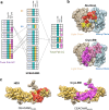
Fig. 3. Comparison of the epitope-paratope interface between hCEACAM5A3-B3 and tusa Fab identified by 3 different structural analysis techniques.
a Comparison of interacting residues in hCEACAM5A3-B3 complexed with tusa Fab. Interacting residues in hCEACAM5 (A3 domain, residues in orange text; B3 domain, residues in blue text) and tusa Fab (heavy chain, residues in purple text; light chain, residues in green text) that were identified by cryo-EM. A subset of these residues was also identified by HDX (yellow) and/or surface plasmon resonance analysis of alanine variants (Ala+SPR, magenta). Not all residues involved in epitopes or paratopes that were identified by HDX or alanine mutagenesis are shown. Hydrogen bonds are depicted by dashed lines. b Comparison of tusa Fab paratope residues identified by alanine mutagenesis (yellow highlighting indicating partial binding and red highlighting indicating no binding) and cryo-EM (light pink highlighting indicating the interacting residues of the heavy chain and magenta highlighting indicating the interacting residues from the light chain). The solvent-accessible surface of the tusa Fab heavy and light chains is illustrated as light brown and light blue, respectively. c Comparison of hCEACAM5 epitope residues identified by HDX (highlighted in red) and cryo-EM (highlighted in magenta). The solvent-accessible surface of hCEACAM5A3-B3 and N-linked glycans are depicted as golden brown and light brown, respectively. b, c Secondary structures (darker ribbons and ball-and-stick moieties) are shown. Ala-Scan, alanine scanning mutagenesis; Ala+SPR, surface plasmon resonance of alanine variants; cryo-EM, cryogenic electron microscopy; HC, heavy chain; hCEACAM5, human carcinoembryonic antigen-related cell adhesion molecule 5; hCEACAM5A3-B3, A3-B3 domains of human carcinoembryonic antigen-related cell adhesion molecule 5; HDX, hydrogen-deuterium exchange; LC, light chain; MAN, mannose; tusa Fab, antigen-binding fragment of tusamitamab.

References

    1. Hammarström, S. The carcinoembryonic antigen (CEA) family: structures, suggested functions and expression in normal and malignant tissues. Semin. Cancer Biol.9, 67–81 (1999). - PubMed
    1. Beauchemin, N. & Arabzadeh, A. Carcinoembryonic antigen-related cell adhesion molecules (CEACAMs) in cancer progression and metastasis. Cancer Metastasis Rev32, 643–671 (2013). - PubMed
    1. Decary, S. et al. Preclinical activity of SAR408701: a novel anti-CEACAM5-maytansinoid antibody-drug conjugate for the treatment of CEACAM5-positive epithelial tumors. Clin. Cancer Res.26, 6589–6599 (2020). - PubMed
    1. Bork, P., Holm, L. & Sander, C. The immunoglobulin fold. Structural classification, sequence patterns and common core. J. Mol. Biol.242, 309–320 (1994). - PubMed
    1. Beauchemin, N., Benchimol, S., Cournoyer, D., Fuks, A. & Stanners, C. P. Isolation and characterization of full-length functional cDNA clones for human carcinoembryonic antigen. Mol. Cell Biol.7, 3221–3230 (1987). - PMC - PubMed

Publication types

MeSH terms

LinkOut - more resources