. 2024 Oct 31;15(1):9407.
doi: 10.1038/s41467-024-53811-3.

Structural basis for human OGG1 processing 8-oxodGuo within nucleosome core particles

Affiliations

Structural basis for human OGG1 processing 8-oxodGuo within nucleosome core particles

Mengtian Ren et al. Nat Commun. .

Abstract

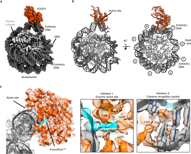
Base excision repair (BER) is initialized by DNA glycosylases, which recognize and flip damaged bases out of the DNA duplex into the enzymes active site, followed by cleavage of the glycosidic bond. Recent studies have revealed that all types of DNA glycosylases repair base lesions less efficiently within nucleosomes, and their repair activity is highly depended on the lesion's location within the nucleosome. To reveal the underlying molecular mechanism of this phenomenon, we determine the 3.1 Å cryo-EM structure of human 8-oxoguanine-DNA glycosylase 1 (hOGG1) bound to a nucleosome core particle (NCP) containing a common oxidative base lesion, 8-oxo-7,8-dihydro-2'-deoxyguanosine (8-oxodGuo). Our structural analysis shows that hOGG1 can recognize and flip 8-oxodGuo even within NCPs; however, the interaction between 8-oxodGuo and hOGG1 in a NCP context is weaker than in free DNA due to competition for nucleosomal DNA by the histones. Binding of OGG1 and the flipping of 8-oxodGuo by hOGG1 leads to a partial detachment of DNA from the histone core and a ratchet-like inward movement of nucleosomal DNA. Our findings provide insights into how the dynamic structure of nucleosomes modulate the activity of repair enzymes within chromatin.


Conflict of interest statement

The authors declare no competing interests.

Figures

Fig. 1
Fig. 1. Impact of 8-oxodGuo location on specific binding by hOGG1K249Q.
a X-ray crystal structure of a NCP (PDB: 3lz0) showing the locations of the tested 8-oxodGuo. The 8-oxodGuo modifications are represented by means of the red sphere model. b 10% native PAGE depicting the bound of hOGG1K249Q with NCP-8-oxodGuo-137. The red arrow highlights a band corresponding to a 1:1 complex of hOGG1K249Q/NCP-8-oxodGuo-137. c Quantification with fits for the binding of hOGG1K249Q to dsDNA. d Quantification with fits for the binding of hOGG1K249Q to NCPs. The presented data display the individual data points of three independent experiments. NCP nucleosome core particle, SHL superhelical location. Source data are provided as a Source Data file.
Fig. 2
Fig. 2. Cryo-EM structure of the hOGG1K249Q/NCP-8-oxodGuo-137 complex.
a DeepEM-enhanced post-processed cryo-EM reconstruction of hOGG1K249Q bound to NCP-8-oxodGuo-137 at 3.1 Å resolution. Light gray: histone octamer, dark gray: DNA, orange: hOGG1. b Side and top views of the derived structural model (ribbon representation) illustrating hOGG1 binding to the 8-oxodGuo lesion without direct interactions with the histone core. c Side view of the structural model with close-up views on the hOGG1 active site overlaid by the density map. Interface I demonstrates interactions between hOGG1K249Q and the 8-oxodGuo lesion in the enzyme’s catalytic pocket. Interface II reveals interactions in the cytosine recognition pocket. Cyan represents 8-oxodGuo-137, dark gray corresponds to DNA, and orange represents hOGG1. Nucleotides and interacting residues are depicted as sticks with color-coded oxygens (red), sulfurs (yellow) and nitrogens (blue).
Fig. 3
Fig. 3. Distortion of the DNA structure upon hOGG1K249Q binding to the NCP-8-oxodGuo-137.
a Alignment of the hOGG1K249Q/NCP-8-oxodGuo-137 complex structure with that of a canonical NCP (PDB: 3lz0). The close-up view highlights the pronounced widening of the minor groove upon hOGG1 binding and the resulting ejection of 8-oxodGuo-137 from the DNA helix. b DNA registry shifts at SHL 5.5 to SHL 7.0 due to local distortions of nucleosomal DNA upon hOGG1 binding. In comparison to a canonical NCP model (PDB: 3lz0), the DNA is pushed inwards from the nearby DNA entry/exit site toward the dyad axis and partially lifted off the NCP core. c Distance plot depicting the minor groove width of the nucleosomal DNA segment spanning from SHL 5.5 to SHL 7.0. The minor groove width was measured between DNA backbone phosphates. Light gray: histone octamer of hOGG1K249Q/NCP-8-oxodGuo-137 complex, dark gray: DNA of hOGG1K249Q/NCP-8-oxodGuo-137 complex, light blue: histone octamer of canonical NCP, dark blue: DNA of canonical NCP, orange: hOGG1. NCP nucleosome core particle. Source data are provided as a Source Data file.
Fig. 4
Fig. 4. Multiple hOGG1 molecules binding to a single dsDNA stretch.
a 2D classes depicting particles with multiple hOGG1K249Q molecules binding to a dsDNA molecule in 2:1 and 3:1 ratios. b Selected 2D class averages of particles displaying a distinctive kinking pattern at each hOGG1K249Q binding site on the dsDNA stretch. c Top view of an in-silico model illustrating six hOGG1 molecules bound to a single free 601 DNA stretch in a random distribution. The model was generated utilizing the crystal structure of hOGG1 bound to free dsDNA (PDB: 1ebm) and the 601 DNA sequence through the x3dna webserver. (http://web.x3dna.org/). Gray: DNA, orange: hOGG1.
Fig. 5
Fig. 5. Two hOGG1 molecules binding to a single NCP-8-oxodGuo-137.
a Illustration of representative 2D classification. b Crude map depicting the binding of hOGG1K249Q in conjunction with NCP-8-oxodGuo-137 at a ratio of 2:1, obtained from the same data collection as presented in Fig. 2. Orange represents hOGG1K249Q bound on 8-oxodGuo-137 (SHL 6.0), Pink corresponds to hOGG1K249Q bound at SHL −2.0. NCP, nucleosome core particle. SHL, superhelical location.

References

    1. Cadet, J. & Davies, K. J. A. Oxidative DNA damage & repair: an introduction. Free Radic. Biol. Med.107, 2–12 (2017). - PMC - PubMed
    1. Poetsch, A. R. The genomics of oxidative DNA damage, repair, and resulting mutagenesis. Comput. Struct. Biotechnol. J.18, 207–219 (2020). - PMC - PubMed
    1. Burrows, C. J. & Muller, J. G. Oxidative nucleobase modifications leading to strand scission. Chem. Rev.98, 1109–1151 (1998). - PubMed
    1. Cadet, J., Douki, T. & Ravanat, J. L. Oxidatively generated damage to the guanine moiety of DNA: mechanistic aspects and formation in cells. Acc. Chem. Res.41, 1075–1083 (2008). - PubMed
    1. Kouchakdjian, M. et al. NMR structural studies of the ionizing radiation adduct 7-hydro-8-oxodeoxyguanosine (8-oxo-7H-dG) opposite deoxyadenosine in a DNA duplex. 8-oxo-7H-dG(syn).dA(anti) alignment at lesion site. Biochemistry30, 1403–1412 (1991). - PubMed

Publication types

LinkOut - more resources