

Handheld multiphoton and pinhole-free reflectance confocal microscopy enables noninvasive, real-time cross-sectional imaging in skin
- PMID: 39478114
- PMCID: PMC11526003
- DOI: 10.1038/s41598-024-76908-7
Handheld multiphoton and pinhole-free reflectance confocal microscopy enables noninvasive, real-time cross-sectional imaging in skin
Abstract
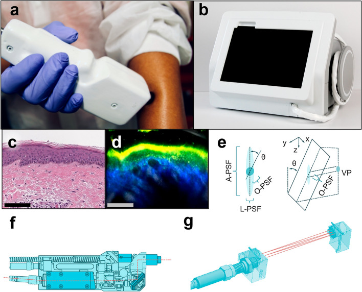
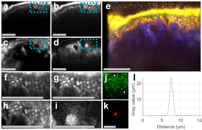
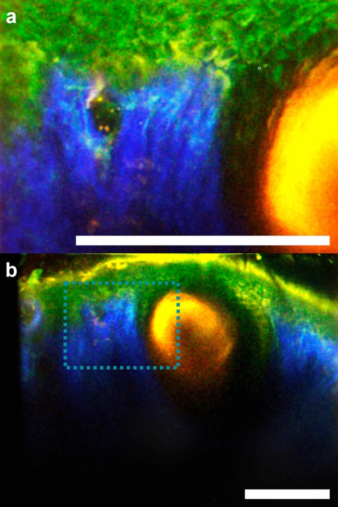
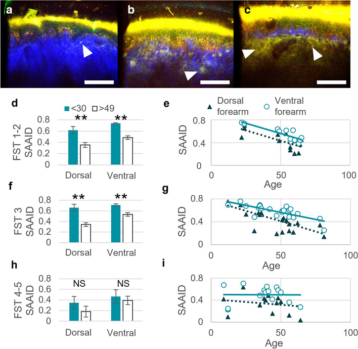
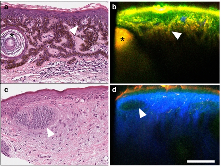
Biopsy-based histology has been the foundation of disease diagnosis and management for over a century. A long-sought goal in dermatology is the development of an imaging modality with sufficient resolution and compositional detail to noninvasively interrogate skin histology in vivo. Here, we describe a system that achieves this goal using cross-sectionally scanned, multimodal microscopy (cross-modal). Cross-modal combines multiphoton and reflectance confocal microscopy into one compact system with coordinated three-axis scanning that preserves optical resolution in cross-section. A custom pinhole-free mechanism employing finite-infinite conjugates further simplifies and stabilizes confocal alignment. Evaluated in participants ages 9-81 and Fitzpatrick skin types (FST) 1-5, cross-modal images revealed histological details analogous to those obtained from traditional biopsied tissue. We observed dermal elastosis in sun-damaged skin, elevated melanin in pigmented skin, basaloid nests in basal cell carcinoma, and elongated rete ridges in seborrheic keratosis, supporting cross-modal's potential to deliver histological insights noninvasively.
© 2024. The Author(s).
Conflict of interest statement
The authors declare the following competing interests: current employment at (K.M., G.S.) and personal financial interest in Enspectra Health (R.N., J.K.).
Figures
Similar articles
-
Role of In Vivo Reflectance Confocal Microscopy in the Analysis of Melanocytic Lesions.Acta Dermatovenerol Croat. 2018 Apr;26(1):64-67. Acta Dermatovenerol Croat. 2018. PMID: 29782304 Review.
-
Research Techniques Made Simple: Emerging Imaging Technologies for Noninvasive Optical Biopsy of Human Skin.J Invest Dermatol. 2022 May;142(5):1243-1252.e1. doi: 10.1016/j.jid.2022.01.016. J Invest Dermatol. 2022. PMID: 35461534 Free PMC article.
-
The challenge of diagnosing seborrheic keratosis by reflectance confocal microscopy.Skin Res Technol. 2018 Nov;24(4):663-666. doi: 10.1111/srt.12582. Epub 2018 May 24. Skin Res Technol. 2018. PMID: 29797357
-
High-resolution imaging of basal cell carcinoma: a comparison between multiphoton microscopy with fluorescence lifetime imaging and reflectance confocal microscopy.Skin Res Technol. 2013 Feb;19(1):e433-43. doi: 10.1111/j.1600-0846.2012.00661.x. Epub 2012 Sep 12. Skin Res Technol. 2013. PMID: 22970856
-
Application of Handheld Confocal Microscopy for Skin Cancer Diagnosis: Advantages and Limitations Compared with the Wide-Probe Confocal.Dermatol Clin. 2016 Oct;34(4):469-475. doi: 10.1016/j.det.2016.05.009. Dermatol Clin. 2016. PMID: 27692452 Review.
Cited by
-
Motion-tolerant 3D volumetric multimodality microscopy imaging of human skin with subcellular resolution and extended field-of-view.Commun Biol. 2025 Feb 5;8(1):186. doi: 10.1038/s42003-025-07614-x. Commun Biol. 2025. PMID: 39910179 Free PMC article.
-
Noninvasive Multimodal Imaging and Its Role in Diagnosing Skin Lesions in Dermatology: A Systematic Review and Meta-Analysis.Am J Clin Dermatol. 2025 Jul 8. doi: 10.1007/s40257-025-00958-4. Online ahead of print. Am J Clin Dermatol. 2025. PMID: 40627274
-
AI-assisted identification of nonmelanoma skin cancer structures based on combined line-field confocal optical coherence tomography and confocal Raman microspectroscopy.J Biomed Opt. 2025 Jul;30(7):076008. doi: 10.1117/1.JBO.30.7.076008. Epub 2025 Jul 28. J Biomed Opt. 2025. PMID: 40726594 Free PMC article.
References
-
- Reiter, O. et al. The diagnostic accuracy of dermoscopy for basal cell carcinoma: A systematic review and meta-analysis. J. Am. Acad. Dermatol.80, 1380–1388 (2019). - PubMed
-
- Caroline, G. et al. In vivo evaluation of skin of children with LC-OCT: An objective assessment. J. Eur. Acad. Dermatol. Venereol.10.1111/JDV.19163 (2023). - PubMed
MeSH terms
LinkOut - more resources
Full Text Sources
Miscellaneous
