

Proteomic analysis of plasma total exosomes and placenta-derived exosomes in patients with gestational diabetes mellitus in the first and second trimesters
- PMID: 39478498
- PMCID: PMC11523606
- DOI: 10.1186/s12884-024-06919-9
Proteomic analysis of plasma total exosomes and placenta-derived exosomes in patients with gestational diabetes mellitus in the first and second trimesters
Abstract
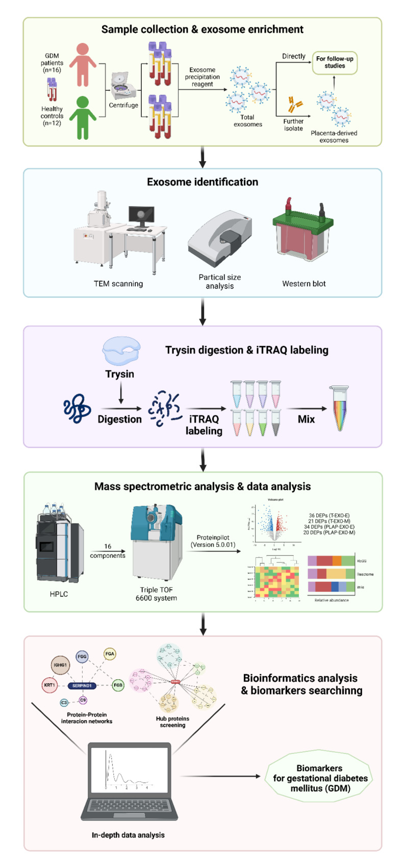
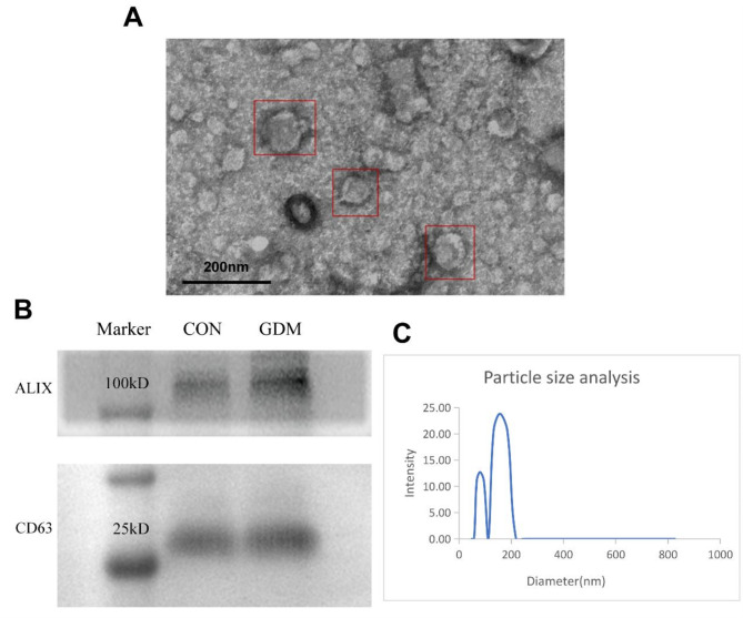
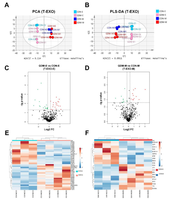
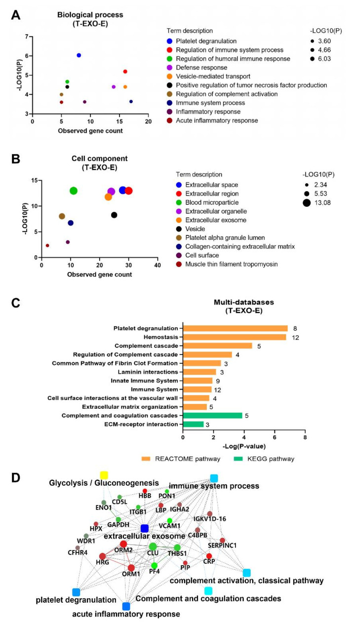
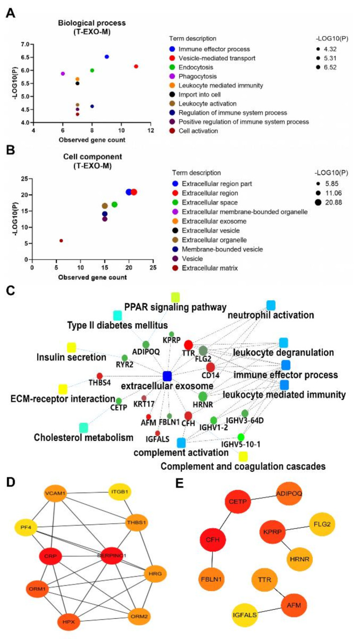
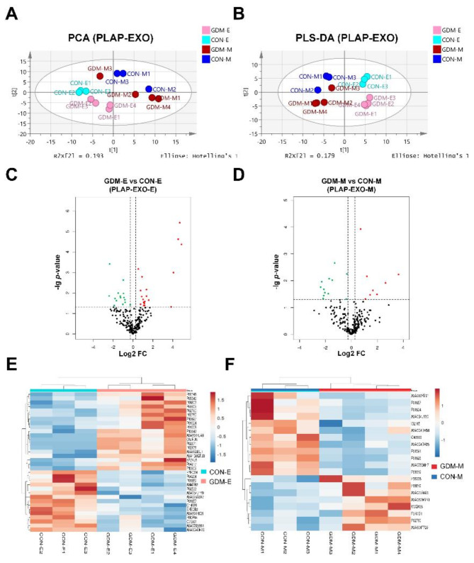
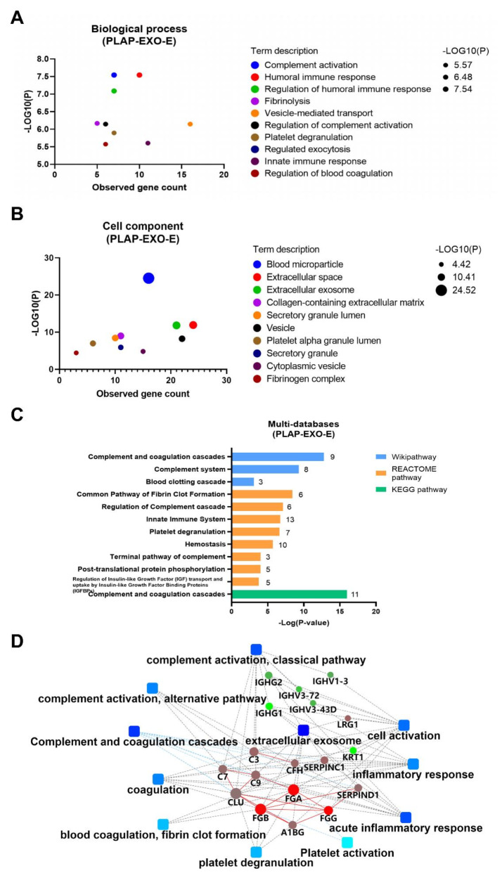
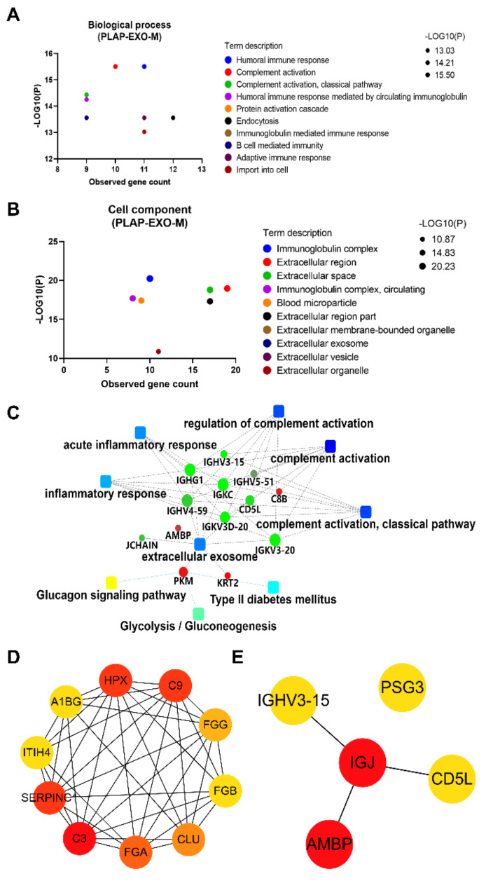
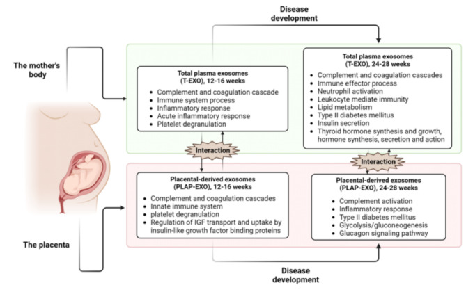
Gestational diabetes mellitus (GDM) is the first spontaneous hyperglycemia during pregnancy. Early diagnosis and intervention are important for the management of the disease. This study compared and analyzed the proteins of total plasma exosomes (T-EXO) and placental-derived exosomes (PLAP-EXO) in pregnant women who subsequently developed GDM (12-16 weeks), GDM patients (24-28 weeks) and their corresponding controls to investigate the pathogenesis and biomarkers of GDM associated with exosomes. The exosomal proteins were extracted and studied by proteomics approach, then bioinformatics analysis was applied to the differentially expressed proteins (DEPs) between the groups. At 12-16 and 24-28 weeks of gestation, 36 and 21 DEPs were identified in T-EXO, while 34 and 20 DEPs were identified in PLAP-EXO between GDM and controls, respectively. These proteins are mainly involved in complement pathways, immunity, inflammation, coagulation and other pathways, most of them have been previously reported as blood or exosomal proteins associated with GDM. The findings suggest that the development of GDM is a progressive process and that early changes promote the development of the disease. Maternal and placental factors play a key role in the pathogenesis of GDM. These proteins especially Hub proteins have the potential to become predictive and diagnostic biomarkers for GDM.
Keywords: Biomarker; Exosome; Gestational diabetes mellitus; Mechanism; Proteomics.
© 2024. The Author(s).
Conflict of interest statement
The authors declare no competing interests.
Figures
Similar articles
-
Early-Pregnancy Serum Maternal and Placenta-Derived Exosomes miRNAs Vary Based on Pancreatic β-Cell Function in GDM.J Clin Endocrinol Metab. 2024 May 17;109(6):1526-1539. doi: 10.1210/clinem/dgad751. J Clin Endocrinol Metab. 2024. PMID: 38127956
-
Gestational Diabetes Mellitus Is Associated With Changes in the Concentration and Bioactivity of Placenta-Derived Exosomes in Maternal Circulation Across Gestation.Diabetes. 2016 Mar;65(3):598-609. doi: 10.2337/db15-0966. Epub 2015 Dec 30. Diabetes. 2016. PMID: 26718504
-
Adipose Tissue Exosomal Proteomic Profile Reveals a Role on Placenta Glucose Metabolism in Gestational Diabetes Mellitus.J Clin Endocrinol Metab. 2019 May 1;104(5):1735-1752. doi: 10.1210/jc.2018-01599. J Clin Endocrinol Metab. 2019. PMID: 30517676
-
Hepatokine levels during the first or early second trimester of pregnancy and the subsequent risk of gestational diabetes mellitus: a systematic review and meta-analysis.Biomarkers. 2021 Sep;26(6):517-531. doi: 10.1080/1354750X.2021.1928754. Epub 2021 Jun 14. Biomarkers. 2021. PMID: 34082623
-
MiRNAs in Gestational Diabetes Mellitus: Potential Mechanisms and Clinical Applications.J Diabetes Res. 2021 Nov 24;2021:4632745. doi: 10.1155/2021/4632745. eCollection 2021. J Diabetes Res. 2021. PMID: 34869778 Free PMC article. Review.
Cited by
-
Long-read transcriptome assembly reveals vast isoform diversity in the placenta associated with metabolic and endocrine function.bioRxiv [Preprint]. 2025 Jun 27:2025.06.26.661362. doi: 10.1101/2025.06.26.661362. bioRxiv. 2025. PMID: 40666916 Free PMC article. Preprint.
References
-
- Association AD. 2. Classification and diagnosis of diabetes: standards of Medical Care in Diabetes—2020. Diabetes Care. 2019;43(Supplement1):S14–31. - PubMed
-
- Federation ID. IDF diabetes atlas. 8th ed. International Diabetes Federation; 2017. pp. 905–911.
MeSH terms
Substances
Grants and funding
LinkOut - more resources
Full Text Sources
