. 2024 Oct 30;23(1):242.
doi: 10.1186/s12943-024-02147-z.

Tumour-intrinsic PDL1 signals regulate the Chk2 DNA damage response in cancer cells and mediate resistance to Chk1 inhibitors

Affiliations

Tumour-intrinsic PDL1 signals regulate the Chk2 DNA damage response in cancer cells and mediate resistance to Chk1 inhibitors

Clare E Murray et al. Mol Cancer. .

Abstract

Background: Aside from the canonical role of PDL1 as a tumour surface-expressed immune checkpoint molecule, tumour-intrinsic PDL1 signals regulate non-canonical immunopathological pathways mediating treatment resistance whose significance, mechanisms, and therapeutic targeting remain incompletely understood. Recent reports implicate tumour-intrinsic PDL1 signals in the DNA damage response (DDR), including promoting homologous recombination DNA damage repair and mRNA stability of DDR proteins, but many mechanistic details remain undefined.

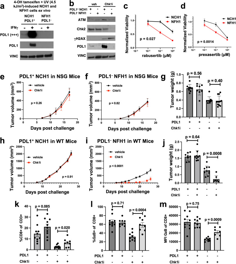
Methods: We genetically depleted PDL1 from transplantable mouse and human cancer cell lines to understand consequences of tumour-intrinsic PDL1 signals in the DNA damage response. We complemented this work with studies of primary human tumours and inducible mouse tumours. We developed novel approaches to show tumour-intrinsic PDL1 signals in specific subcellular locations. We pharmacologically depleted tumour PDL1 in vivo in mouse models with repurposed FDA-approved drugs for proof-of-concept clinical translation studies.

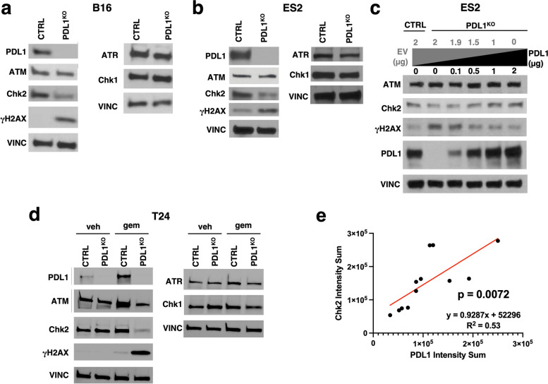
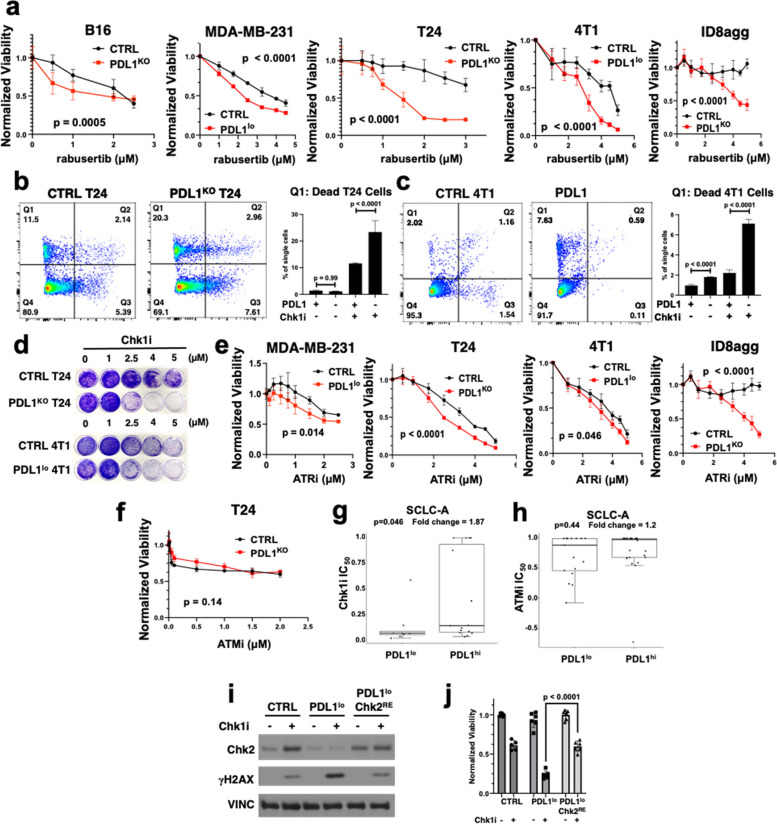
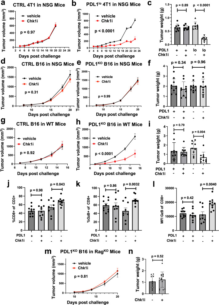
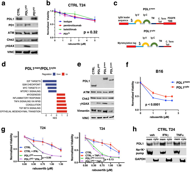
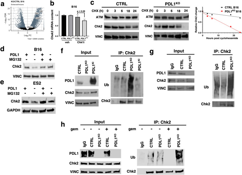
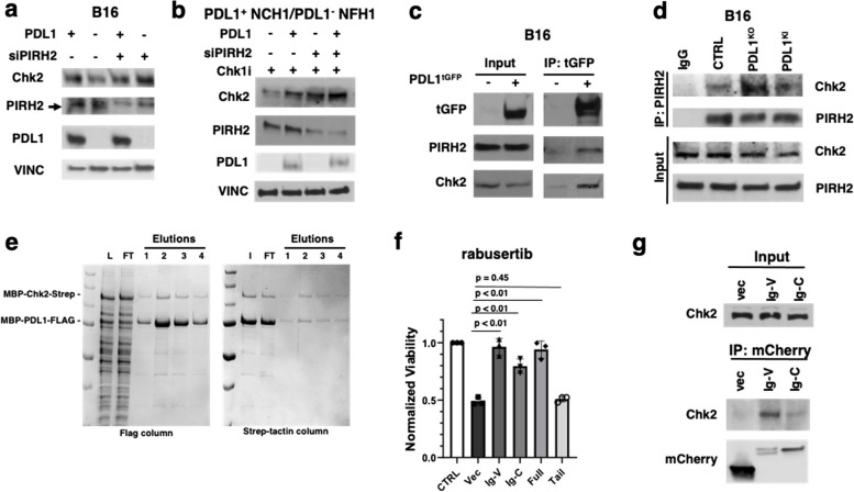
Results: We show that tumour-intrinsic PDL1 promotes the checkpoint kinase-2 (Chk2)-mediated DNA damage response. Intracellular but not surface-expressed PDL1 controlled Chk2 protein content post-translationally and independently of PD1 by antagonising PIRH2 E3 ligase-mediated Chk2 polyubiquitination and protein degradation. Genetic tumour PDL1 depletion specifically reduced tumour Chk2 content but not ATM, ATR, or Chk1 DDR proteins, enhanced Chk1 inhibitor (Chk1i) synthetic lethality in vitro in diverse human and murine tumour models, and improved Chk1i efficacy in vivo. Pharmacologic tumour PDL1 depletion with cefepime or ceftazidime replicated genetic tumour PDL1 depletion by reducing tumour Chk2, inducing Chk1i synthetic lethality in a tumour PDL1-dependent manner, and reducing in vivo tumour growth when combined with Chk1i.

Conclusions: Our data challenge the prevailing surface PDL1 paradigm, elucidate important and previously unappreciated roles for tumour-intrinsic PDL1 in regulating the ATM/Chk2 DNA damage response axis and E3 ligase-mediated protein degradation, suggest tumour PDL1 as a biomarker for Chk1i efficacy, and support the rapid clinical potential of pharmacologic tumour PDL1 depletion to treat selected cancers.

Keywords: Chk2; DDR inhibitors; DNA damage repair; Immune checkpoints; PDL1; Synthetic lethality.


Conflict of interest statement

TJC and AVK have filed a patent on using PDL1 depleting drugs to treat cancer.

Figures

Fig. 1
Fig. 1
Tumour PDL1 promotes Chk2 content. a. Immunoblot from whole cell lysates of CTRL and PDL1KO B16 cells. Vinculin (VINC), loading control. γH2AXSer139 blot is shown as γH2AX. b. Immunoblot from whole cell lysates of CTRL and PDL1KO ES2 cells. VINC, loading control. c. Immunoblot of CTRL and PDL1KO ES2 cells transfected with empty vector (EV, gray) and/or PDL1 plasmid (black) at depicted μg. d. Immunoblot of CTRL versus PDL1KO T24 cells after 72-h incubation with vehicle or gemcitabine (5 ng/mL). e. Quantification of relative intracellular PDL1 and Chk2 expression of 12 human high-grade serous ovarian tumours at upfront treatment. Statistical analysis by linear regression
Fig. 2
Fig. 2
Tumour PDL1 deficiency promotes Chk2-dependent synthetic lethality to Chk1i and ATRi in vitro. a. In vitro cell viability of CTRL versus PDL1KO B16, T24, and ID8agg cells or CTRL versus PDL1lo 4T1 and MDA-MB-231 cells incubated with the selective Chk1i rabusertib for 5 days at indicated doses (mean ± SD, n = 3 independent replicates, P values by two-way ANOVA). Viability normalised to vehicle controls. b. Flow cytometry analysis of Annexin/PI-stained CTRL versus PDL1KO T24 cells and c. CTRL versus PDL1lo 4T1 cells after treatment with vehicle (DMSO) or 1 µM rabusertib. Q1 (% dead cells) quantified. P, two-way ANOVA. d. Clonogenic crystal violet stain of PDL1KO T24 or PDL1lo 4T1 and respective CTRL cells treated with indicated rabusertib (Chk1i) concentrations for 5 days. e. Cell viability of indicated CTRL versus PDL1-deficient cell lines after ATRi (AZD6738) or f. ATMi (AZD0156) incubation. P, two-way ANOVA. g. IC50 for the Chk1i prexasertib and h. ATMi AZD0156 in 44 human SCLC-A cell lines segregated by low (lo) or high (hi) PDL1 expression. P, two-sided t-test. Grubb’s test was used to identify outliers. i. Immunoblot after Chk2 re-expression plasmids (Chk2RE) or empty vector controls were transiently introduced into CTRL or PDL1lo 4T1 cells and treated with rabusertib (Chk1i, 2.5 µM) or vehicle (DMSO) for 72 h. j. PDL1lo 4T1 viability with transient Chk2RE as in i, treated with 2.5 µM rabusertib for 5 days in vitro versus CTRL and PDL1lo with empty vector. Data are mean ± SD normalised to vehicle. P, unpaired t-test
Fig. 3
Fig. 3
Tumour PDL1 depletion promotes Chk1i synthetic lethality in vivo that requires adaptive immunity in distinct models. Growth in mammary fat pad of a. CTRL and b. PDL1lo 4T1 tumours in immune deficient NSG mice treated with vehicle or rabusertib (Chk1i). Data represented as mean ± SEM. P by two-way ANOVA. c. Tumour end-point weights from a and b. P, unpaired t-test. d-e. Tumour growth curves of NSG mice challenged with d. CTRL and e. PDL1KO B16 cells and treated with rabusertib daily. P, two-way ANOVA. f. NSG mice bearing B16 tumours as in d-e were sacrificed day 21 post-challenge and tumours were weighed. P, unpaired t-test. g-h. Growth of subcutaneous g. CTRL versus h. PDL1KO B16 tumours in WT mice treated with vehicle or rabusertib. Data represented as mean ± SEM. P, two-way ANOVA. i. Tumour end-point weights from g and h. P, unpaired t-test. j-l. Flow cytometry immune analyses of indicated populations in CTRL versus PDL1KO B16 tumours treated with vehicle or rabusertib in vivo. Mice were sacrificed day 17 post-challenge. P, unpaired t-test. m. Tumour growth curves of Rag2KO mice bearing PDL1KO B16 tumours treated with vehicle or rabusertib daily. P, two-way ANOVA. n. Rag2KO mice as in m were sacrificed day 20 post-challenge. P, unpaired t-test
Fig. 4
Fig. 4
Autochthonous PDL1-deficient tumour cells phenocopy genetic PDL1 silencing by inducing Chk2 deficiency and Chk1i synthetic lethality in vitro and in vivo. a. Immunoblot validation of PDL1 expression in NRASQ61R-mutant melanoma cells derived from CTRL TN61R (NCH1; PDL1+) or PDL1 TN61R (NFH1; PDL1) mice. Cells were treated in vitro with 0.1 ng/mL IFNγ for 48 h. b. Immunoblot of NCH1 (PDL1+) or NFH1 (PDL1) cells treated in vitro with the Chk1i rabusertib or vehicle (veh) for indicated proteins. c-d. Cell viability of PDL1+ NCH1 versus PDL1 NFH1 melanoma cells treated with the Chk1i c. rabusertib or d. prexasertib for 72 h in vitro. Viability normalised to vehicle. P, two-way ANOVA. NSG mice were challenged with e. PDL1+ NCH1 cells or f. PDL1 NFH1 cells and treated with rabusertib daily. P, two-way ANOVA. g. Tumour-bearing NSG mice were sacrificed day 18 post-challenge. Tumours were excised and weighed. P, unpaired t-test. WT mice were challenged with h. NCH1 or i. NFH1 cells and treated with vehicle or rabusertib daily. P, two-way ANOVA. j. Tumour-bearing WT mice were sacrificed, and tumours were excised and weighed. P, unpaired t-test. k-m. WT mice bearing NCH1 or NFH1 tumours were sacrificed day 28 post-challenge and profiled for indicated immune populations by flow cytometry. MFI, mean fluorescence intensity. P, unpaired t-test
Fig. 5
Fig. 5
Tumour PDL1 control of Chk2 is through intracellular PDL1 and independent of PD1 and immunity. a. Immunoblots of PDL1KO and PD1KO B16 cells versus CTRL B16 cells for indicated proteins. Vinculin (VINC) loading control. b. In vitro MTT cell viability of CTRL T24 cells treated with the αPD1 antibodies pembrolizumab (50 µg/mL), balstilimab (50 µg/mL), or genetic shRNA PD1 knockdown (PD1lo) with increasing doses of the Chk1i rabusertib for 5 days at indicated doses. P by two-way ANOVA. Viability shown as relative values following normalisation to respective isotype or vehicle-treated controls. c. Schematic of membrane (mem)-localising versus intracellular (cyto)-localising PDL1 expression constructs (see methods). d. RNA-seq analyses of PDL1mem versus PDL1cyto B16 cells using gene set enrichment analysis. Top 10 altered hallmark pathways (p < 0.01) are shown by PDL1 subcellular location. e. Immunoblot comparing ATM, Chk2, γH2AX, and vimentin expression of indicated B16 cells. Vinculin (VINC) as loading control. f. MTT viability assay of PDL1mem versus PDL1cyto cells after 96-h of in vitro treatment with the Chk1i rabusertib at indicated concentrations. P, two-way ANOVA. g. Viability analysis of CTRL versus PDL1KO T24 cells treated with indicated rabusertib concentrations ± 1 ng/mL IFNγ, 10 ng/mL TNFα, or vehicle. P, two-way ANOVA. h. Immunoblot of cell lysates of membrane and cytosolic fractions of CTRL T24 cells treated with vehicle (PBS), IFNγ, or TNFα as in g. Na+/K+ pump as membrane loading control and GAPDH as cytosolic loading control.
Fig. 6
Fig. 6
PDL1 protects Chk2 from ubiquitination in distinct tumours. a. Volcano plot of RNA-seq data depicting PDL1KO effect on select mRNA levels versus CTRL B16. b. qRT-PCR of Chek2 mRNA in CTRL versus PDL1KO B16 cells treated with DMSO or rabusertib. P, unpaired t-test. c. Immunoblot of CTRL versus PDL1KO B16 cells treated with cycloheximide (CHX). Summary graph depicts normalised total Chk2 protein levels quantified using ImageJ. d. Immunoblot of CTRL versus PDL1KO B16 and e. ES2 cells treated with MG132 (2 µM) for 12-h. f. Immunoblot of immunoprecipitated Chk2 from CTRL, PDL1KO, and stable PDL1 knock-in (PDL1KI) B16 cells and g. CTRL and PDL1KO ES2 cells after MG132 treatment. h. CTRL and PDL1KO T24 cells treated with MG132 for 16-h plus vehicle (H2O) or 10 ng/mL gemcitabine (gem). Endogenous Chk2 was immunoprecipitated and Chk2 and ubiquitination (Ub) were detected by immunoblot
Fig. 7
Fig. 7
Tumour PDL1 protects Chk2 from PIRH2 E3 ligase-mediated degradation in distinct tumours through its immunoglobulin-like domains. a. Non-targeting or PIRH2-specific siRNA vectors transfected into CTRL or PDL1KO B16 cells or b. PDL1+ NCH1 cells and PDL1 NFH1 cells. c. Interaction among PDL1, Chk2 and PIRH2 in B16 cells stably expressing vector control or turbo-GFP (tGFP)-tagged PDL1 (PDL1tGFP) treated with MG132 and co-immunoprecipitated with tGFP trap beads followed by immunoblotting. d. Endogenous PIRH2 co-immunoprecipitated in CTRL, PDL1KO, and PDL1KI B16 cells. e. Left: Flag-tagged human PDL1 and Streptavidin-tagged human Chk2 were co-expressed in Tni cells and affinity pull-downs of lysates with Flag resin and eluate fractions (1,2,3,4) followed by SDS-PAGE and Coomassie blue staining. Right: eluates were pooled for reciprocal Chk2 affinity pulldown using Strep-tactin resin. L, lysates; FT, flow-through; I, input. f. Viability of PDL1 truncation mutant-expressing B16 cells 72-h post rabusertib (2.5 μM) treatment. P, unpaired t-test. g. Immunoblot of domain-expressing B16 cells immunoprecipitated using anti-mCherry beads
Fig. 8
Fig. 8
Pharmacologic tumour PDL1 depletion replicates genetic tumour PDL1KO by depleting Chk2 and inducing Chk1i synthetic lethality. a. Immunoblot of CTRL T24 cells for indicated proteins after 72 h of treatment with vehicle (DMSO), 1 µM rabusertib (Chk1i), 80 μM cefepime, or combination. b. Immunoblot of CTRL, PDL1KO, and stable PDL1KI B16 cells treated with 80 μM cefepime for indicated time points. c-e. Cell viability of c. CTRL MDA-MB-231, d. CTRL T24, and e. PDL1KO T24 cells treated with indicated concentrations of rabusertib ± 80 μM cefepime or vehicle (DMSO) for 96 h. P, two-way ANOVA. f. Immunoblot of CTRL T24 cells treated with vehicle, 1 μM rabusertib, 80 μM ceftazidime, or both for 72 h. g. Immunoblot of CTRL, PDL1KO and stable PDL1KI B16 cells treated with 80 μM ceftazidime for indicated time points. h-j. Cell viability of h. CTRL MDA-MB-231, i. CTRL T24, and j. PDL1KO T24 cells treated with indicated concentrations of rabusertib ± 80 μM ceftazidime or vehicle for 96 h. P, two-way ANOVA. k. MTT viability analysis of CTRL T24 cells treated for 96 h with 80 μM of indicated β-lactams with rabusertib. P, two-way ANOVA. l. Growth curves of PDL1+ NCH1 tumours implanted subcutaneously in WT mice treated with vehicle, ceftazidime, rabusertib, or both. P, two-way ANOVA. m. Tumour weights of NCH1 tumours from l. at endpoint (day 18). P, unpaired t-test. Vehicle (veh), ceftazidime (ctz), rabusertib (rab), and ceftazidime + rabusertib (combo) groups are indicated

References

    1. Topalian SL, Taube JM, Anders RA, Pardoll DM. Mechanism-driven biomarkers to guide immune checkpoint blockade in cancer therapy. Nat Rev Cancer. 2016;16:275–87. 10.1038/nrc.2016.36. - PMC - PubMed
    1. Taube JM, et al. Colocalisation of inflammatory response with B7–h1 expression in human melanocytic lesions supports an adaptive resistance mechanism of immune escape. Sci Transl Med. 2012;4:127ra137. 10.1126/scitranslmed.3003689 https://doi.org:4/127/127ra37 . - PMC - PubMed
    1. Topalian, S. L., Drake, C. G. & Pardoll, D. M. J. C. o. i. i. Targeting the PD-1/B7-H1 (PD-L1) pathway to activate anti-tumour immunity. 24, 207–212 (2012). - PMC - PubMed
    1. Paterson AM, et al. The programmed death-1 ligand 1:b7–1 pathway restrains diabetogenic effector T cells in vivo. J Immunol. 2011;187:1097–105. 10.4049/jimmunol.1003496 https://doi.org:jimmunol.1003496 . - PMC - PubMed
    1. Dong H, et al. tumour-associated B7–H1 promotes T-cell apoptosis: A potential mechanism of immune evasion. Nat Med. 2002;8:793–800. - PubMed

LinkOut - more resources