. 2024 Oct 30;28(1):350.
doi: 10.1186/s13054-024-05127-3.

Airway epithelium damage in acute respiratory distress syndrome

Affiliations

Airway epithelium damage in acute respiratory distress syndrome

Ludovic Gerard et al. Crit Care. .

Abstract

Background: The airway epithelium (AE) fulfils multiple functions to maintain pulmonary homeostasis, among which ensuring adequate barrier function, cell differentiation and polarization, and actively transporting immunoglobulin A (IgA), the predominant mucosal immunoglobulin in the airway lumen, via the polymeric immunoglobulin receptor (pIgR). Morphological changes of the airways have been reported in ARDS, while their detailed features, impact for mucosal immunity, and causative mechanisms remain unclear. Therefore, this study aimed to assess epithelial alterations in the distal airways of patients with ARDS.

Methods: We retrospectively analyzed lung tissue samples from ARDS patients and controls to investigate and quantify structural and functional changes in the small airways, using multiplex fluorescence immunostaining and computer-assisted quantification on whole tissue sections. Additionally, we measured markers of mucosal immunity, IgA and pIgR, alongside with other epithelial markers, in the serum and the broncho-alveolar lavage fluid (BALF) prospectively collected from ARDS patients and controls.

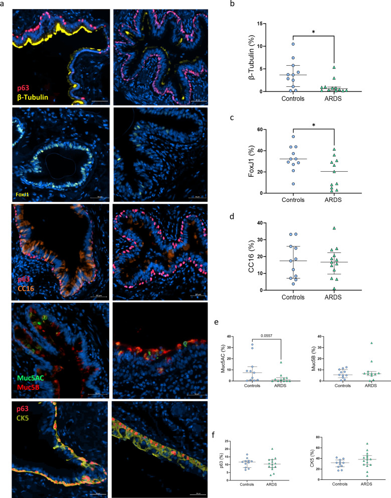
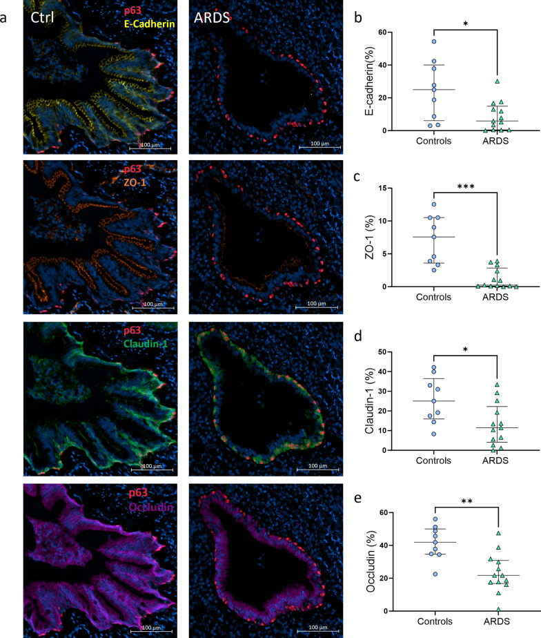
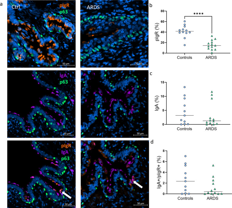
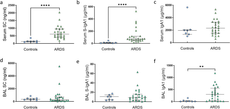
Results: Compared to controls, airways of ARDS were characterized by increased epithelial denudation (p = 0.0003) and diffuse epithelial infiltration by neutrophils (p = 0.0005). Quantitative evaluation of multiplex fluorescence immunostaining revealed a loss of ciliated cells (p = 0.0317) a trend towards decreased goblet cells (p = 0.056), and no change regarding cell progenitors (basal and club cells), indicating altered mucociliary differentiation. Increased epithelial permeability was also shown in ARDS with a significant decrease of tight (p < 0.0001) and adherens (p = 0.025) junctional proteins. Additionally, we observed a significant decrease of the expression of pIgR, (p < 0.0001), indicating impaired mucosal IgA immunity. Serum concentrations of secretory component (SC) and S-IgA were increased in ARDS (both p < 0.0001), along other lung-derived proteins (CC16, SP-D, sRAGE). However, their BALF concentrations remained unchanged, suggesting a spillover of airway and alveolar proteins through a damaged AE.

Conclusion: The airway epithelium from patients with ARDS exhibits multifaceted alterations leading to altered mucociliary differentiation, compromised defense functions and increased permeability with pneumoproteinemia.

Keywords: ARDS; Airway epithelium; Ciliated cells; Immunoglobulin A; Junctional proteins; Mucosal immunity; Pneumoproteinemia.


Conflict of interest statement

The authors declare no competing interests.

Figures

Fig. 1
Fig. 1
Structural changes in the airways of ARDS patients. a, b Representative HE staining of samples from a control (left) and a patient with ARDS (right) showing epithelial denudation (a, white arrow) and neutrophilic infiltration (b). c, d Quantitative analysis comparing the extent of epithelial denudation (c), and the presence of inflammatory cells infiltrating airway epithelia, using a four-grade scale (d) in controls (n = 15) and ARDS (n = 25). Between-group difference was evaluated using the non-parametric Mann–Whitney U test. *** indicates p < 0.0005
Fig. 2
Fig. 2
Changes in airway epithelial populations. a Representative immunofluorescence staining of sections from a control (left) and an ARDS patient (right) stained for β-tubulin and Fox-J1 (ciliated cells), CC16/SCGB1A1 (club cells), Muc5AC and Muc5B (goblet cells) and p63/CK5 (basal cells). Scale bars, 50 µm. be Quantification of the area expressing β-tubulin (in proportion of the total AE area, (b), Fox-J1 (c), CC16/SCGB1A1 (d), Muc5AC and Muc5B (e), p63 and CK5 (f) showing a loss of ciliated cells in patients with ARDS (n = 13), compared to controls (n = 11). Between-group difference was evaluated using the non-parametric Mann–Whitney U test. *indicates p < 0.05
Fig. 3
Fig. 3
Loss of tight and adherens junctions in the airways of patients with ARDS. a Representative immunofluorescence staining of sections from a control (left) and an ARDS patient (right) stained with E-cadherin, Zonula Occludens-1 (ZO-1), Claudin-1 and Occludin, costained with p63 and Hoechst. Scale bars, 100 µm. be Quantification of the area expressing E-Cadherin (in proportion of the total AE area, (b), ZO-1 (c), Claudin-1 (d), and Occludin (e) showing a loss of adherens (E-Cadherin) and tight (ZO-1, Claudin-1, Occludin) junctions in patients with ARDS (n = 13), compared to controls (n = 9). Between group difference was evaluated using the non-parametric Mann–Whitney U test. *indicates p < 0.05, ** indicates p < 0.005 and *** indicates p < 0.0005
Fig. 4
Fig. 4
Decreased pIgR expression in the airways of patients with ARDS. a Representative immunofluorescence staining of sections from a control (left) and an ARDS patient (right) stained for polymeric Immunoglobulin Receptor (pIgR, upper panel), Immunoglobulin A (IgA, middle panel) and both pIgR and IgA (white arrows, lower panel), costained for p63 and Hoechst. Scale bars, 50 µm. bd Quantification of the area expressing pIgR (b), IgA (c), and of the area co-expressing IgA and pIgR (reflecting Secretory IgA [S-IgA], (d), all expressed in proportion of the total AE area, showing a significant decrease of pIgR expression within the AE of ARDS patients (n = 12) compared to controls (n = 13). Between-group difference was evaluated using the non-parametric Mann–Whitney U test. **** indicates p < 0.0001
Fig. 5
Fig. 5
Dysregulated IgA-pIgR/SC system in ARDS patients. ad Serum secretory component (SC, a), and S-IgA1 (b) concentrations are increased in the serum of patients with ARDS (n = 24) compared to controls (n = 7), whereas no difference was found regarding total IgA1 (c). ef Conversely, no difference was found in the BALF concentrations of SC (d), S-IgA (e) and S-IgA1 (f) comparing the same ARDS patients to controls, whereas total IgA1 is increased. Between-group difference was evaluated using the non-parametric Mann–Whitney U test. ** indicates p < 0.005, ****p < 0.0001

References

    1. Bellani G, Laffey JG, Pham T, Fan E, Brochard L, Esteban A, et al. Epidemiology, patterns of care, and mortality for patients with acute respiratory distress syndrome in intensive care units in 50 countries. JAMA. 2016;315(8):788–800. - PubMed
    1. Thompson BT, Chambers RC, Liu KD. Acute respiratory distress syndrome. N Engl J Med. 2017;377(6):562–72. - PubMed
    1. Morales MM, Pires-Neto RC, Inforsato N, Lancas T, da Silva LF, Saldiva PH, et al. Small airway remodeling in acute respiratory distress syndrome: a study in autopsy lung tissue. Crit Care. 2011;15(1):R4. - PMC - PubMed
    1. Jain M, Sznajder JI. Bench-to-bedside review: distal airways in acute respiratory distress syndrome. Crit Care. 2007;11(1):206. - PMC - PubMed
    1. Ortiz G, Garay M, Capelozzi V, Cardinal-Fernandez P. Airway pathological alterations selectively associated with acute respiratory distress syndrome and diffuse alveolar damage: narrative review. Arch Bronconeumol (Engl Ed). 2019;55(1):31–7. - PubMed

Publication types

LinkOut - more resources