

doi: 10.1016/j.eats.2024.103075.
eCollection 2024 Oct.
Microfragmented Adipose Tissue Associated With Collagen Membrane in the Treatment of Focal Knee Cartilage Defect
Affiliations
- PMID: 39479029
- PMCID: PMC11519851
- DOI: 10.1016/j.eats.2024.103075
Item in Clipboard
Microfragmented Adipose Tissue Associated With Collagen Membrane in the Treatment of Focal Knee Cartilage Defect
Arthrosc Tech.
.
Abstract
Focal articular cartilage defects are an important factor that leads to dysfunction of the knee joint. Several different surgical approaches have been tried, most of them showing poor results in the long term. The use of orthobiologics in the context of focal chondral lesion has emerged as a potential tool in the treatment of this condition. In this article, we present a surgical technique for the treatment of focal chondral lesions using a collagen membrane associated with microfragmented adipose tissue graft.
© 2024 The Authors.
Figures
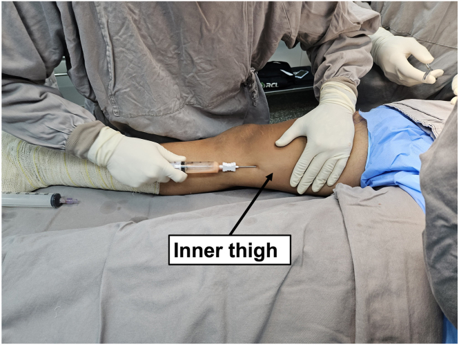
Medial view of the right limb. Inner thigh fat harvesting using a 20-mL VacLok syringe (Merit Medical Systems).
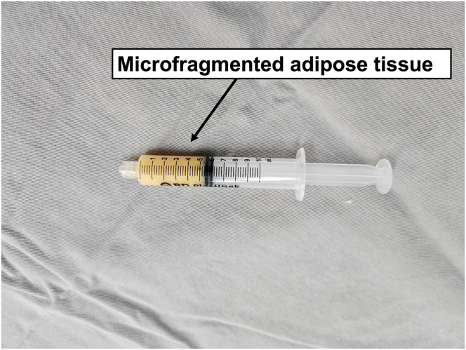
A syringe containing 5 mL microfragmented adipose tissue obtained after Lipogems processing (Lipogems International SpA).
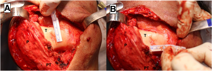
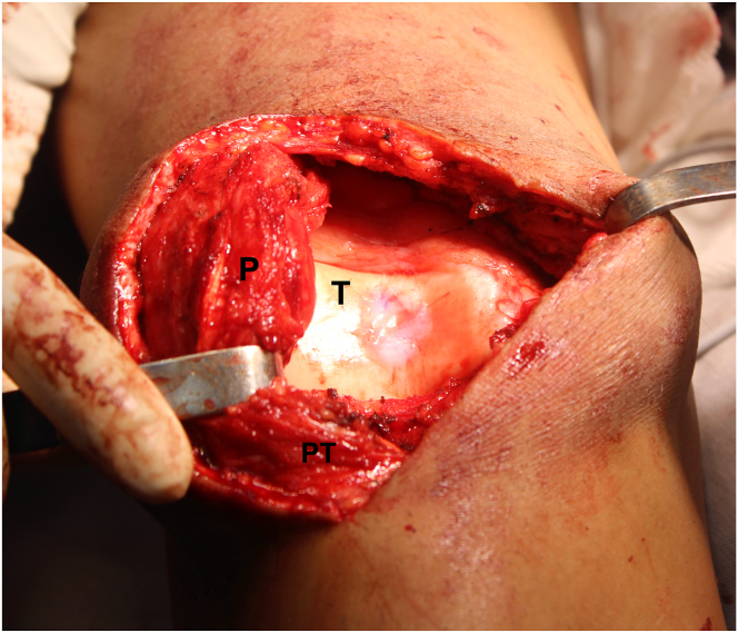
Front view of the right knee. Measurement of the chondral defect on the center of the trochlea on the vertical (A) and horizontal axis (B). (P, patella; PT, patellar tendon; T: trochlea.)
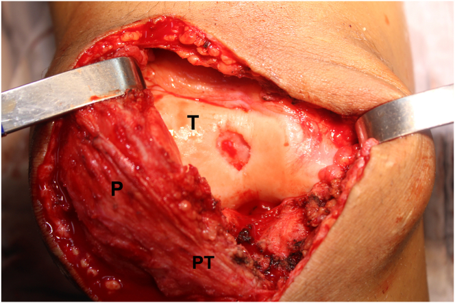
Front view of the right knee. Aspect of the chondral defect on the center of the trochlea after removing residual cartilage fragments from the lesion bed, down to the subchondral bone, and regularizing the edges of the lesion. (P, patella; PT, patellar tendon; T: trochlea.)
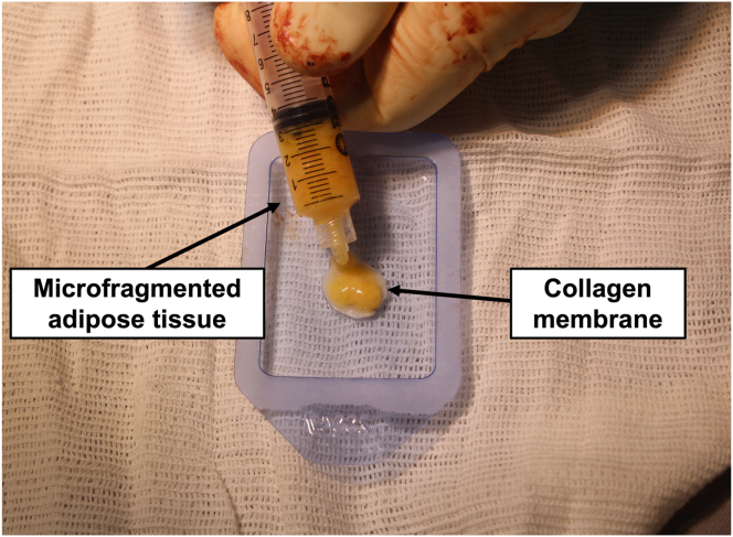
On the auxiliary table, the prepared microfragmented adipose tissue is deposited on the rough surface of the collagen membrane.
Front view of the right knee. The patella is retracted laterally, allowing proper exposure of the trochlea. In the center of the trochlea can be visualized the final aspect of the collagen membrane associated with the microfragmented adipose tissue. (P, patella; PT, patellar tendon; T: trochlea.)
References
-
- Niemeyer P., Albrecht D., Andereya S., et al. Autologous chondrocyte implantation (ACI) for cartilage defects of the knee: A guideline by the working group “Clinical Tissue Regeneration” of the German Society of Orthopaedics and Trauma (DGOU) Knee. 2016;23:426–435. - PubMed
LinkOut - more resources
Full Text Sources
