. 2024 Dec 4;12(47):12307-12319.
doi: 10.1039/d4tb01628a.

Cell-specific spatial profiling of targeted protein expression to characterize the impact of intracortical microelectrode implantation on neuronal health

Affiliations

Cell-specific spatial profiling of targeted protein expression to characterize the impact of intracortical microelectrode implantation on neuronal health

Lindsey N Druschel et al. J Mater Chem B. .

Abstract

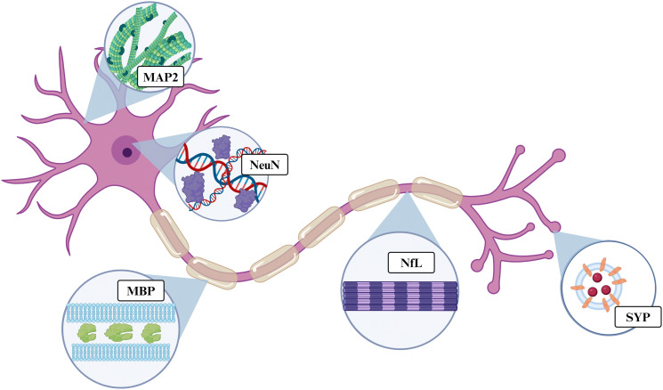
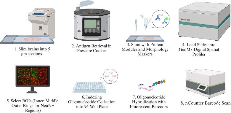
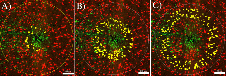
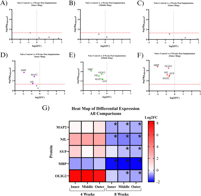
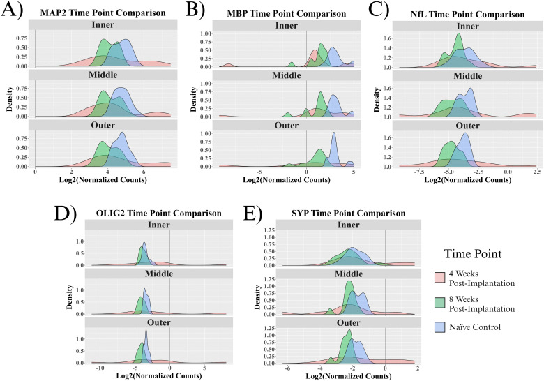
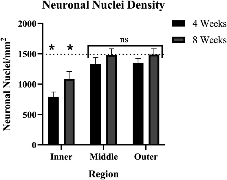
Intracortical microelectrode arrays (MEAs) can record neuronal activity and advance brain-computer interface (BCI) devices. Implantation of the invasive MEA kills local neurons, which has been documented using immunohistochemistry (IHC). Neuronal nuclear protein (NeuN), a protein that lines the nuclei of exclusively neuronal cells, has been used as a marker for neuronal health and survival for decades in neuroscience and neural engineering. NeuN staining is often used to describe the neuronal response to intracortical microelectrode array (MEA) implantation. However, IHC is semiquantitative, relying on intensity readings rather than directly counting expressed proteins. To supplement previous IHC studies, we evaluated the expression of proteins representing different aspects of neuronal structure or function: microtubule-associated protein 2 (MAP2), neurofilament light (NfL), synaptophysin (SYP), myelin basic protein (MBP), and oligodendrocyte transcription factor 2 (OLIG2) following a neural injury caused by intracortical MEA implantation. Together, these five proteins evaluate the cytoskeletal structure, neurotransmitter release, and myelination of neurons. To fully evaluate neuronal health in NeuN-positive (NeuN+) regions, we only quantified protein expression in NeuN+ regions, making this the first-ever cell-specific spatial profiling evaluation of targeted proteins by multiplex immunochemistry following MEA implantation. We performed our protein quantification along with NeuN IHC to compare the results of the two techniques directly. We found that NeuN immunohistochemical analysis does not show the same trends as MAP2, NfL, SYP, MBP, and OLIG2 expression. Further, we found that all five quantified proteins show a decreased expression pattern that aligns more with historic intracortical MEA recording performance.


Conflict of interest statement

The authors declare no conflict of interest.

Figures

Fig. 1
Fig. 1. Simplified illustration of the general location of each protein on a motor neuron. OLIG2 is not illustrated, as it is a signaling protein rather than structural. Created with https://BioRender.com.
Fig. 2
Fig. 2. Illustration of protein quantification methods. Intermediate steps such as washes or fixation are not illustrated but are described at length in the provided protocol.
Fig. 3
Fig. 3. A cross-section of an MEA implant site stained for neuronal nuclei (NeuN, red) and activated astrocytes (GFAP, green), showing the segmented NeuN+ inner ((A), 0–90 μm), middle ((B), 90–180 μm), and outer ((C), 180–270 μm) regions (yellow).
Fig. 4
Fig. 4. Quantification of protein counts. (A)–(C) Volcano plots of protein expression within the NeuN+ region in 4-weeks post-implantation mice compared to the NeuN+ region in naïve control mice. The red line is the significance threshold padj < 0.05. All significant proteins are labeled (only MBP). (D)–(F) Volcano plot of protein expression within the NeuN+ region in 8-weeks post-implantation mice compared to the NeuN+ region in naïve controls. All significant proteins are labeled (all proteins were significant in the middle and outer regions). (A) and (D) Analysis of inner ring only. (B) and (E) Analysis of middle ring only. (C) and (F) Analysis of outer ring only. (G) Heat map of protein expression. This figure combines data from (A)–(F). Black asterisks represent significant differential expression between experimental groups compared to naïve controls. The log2FC scale on the right represents comparisons that have significant differential expression, ranging from low magnitude log2FC values (dark blue) to high magnitude log2FC values (red). This figure was assembled with https://BioRender.com.
Fig. 5
Fig. 5. Probability density functions of measured proteins. The individual probability density functions show the distribution of protein expression between samples for the inner, middle, and outer rings of each protein within experimental and control groups.
Fig. 6
Fig. 6. Bar graph of neuronal nuclei density as a function of time and distance from the implant, showing inner (0–90 μm), middle (90–180 μm), and outer (180–270 μm) rings. Error bars represent the standard error of each group. Asterisks (*) indicate significance (p < 0.02) compared to a baseline of naïve control mice. The baseline neuronal nuclear density is ∼1534 NN mm−2, indicated on this plot with a dotted line.

Similar articles

Cited by

References

    1. Wellman S. M. et al., Aberrant accumulation of age- and disease-associated factors following neural probe implantation in a mouse model of Alzheimer’s disease. J. Neural Eng. 2023;20(4):046044. doi: 10.1088/1741-2552/aceca5. - DOI - PMC - PubMed
    1. Fan C. et al., Plug-and-Play Stability for Intracortical Brain-Computer Interfaces: A One-Year Demonstration of Seamless Brain-to-Text Communication. Adv. Neural Inf. Process. Syst. 2023;36:42258–42270. - PMC - PubMed
    1. Benazzouz A. et al., Intraoperative microrecordings of the subthalamic nucleus in Parkinson's disease. Mov. Disord. 2002;17(Suppl 3):S145–S149. doi: 10.1002/mds.10156. - DOI - PubMed
    1. Rapeaux A. B. Constandinou T. G. Implantable brain machine interfaces: first-in-human studies, technology challenges and trends. Curr. Opin. Biotechnol. 2021;72:102–111. doi: 10.1016/j.copbio.2021.10.001. - DOI - PubMed
    1. Badakva A. M. Miller N. V. Zobova L. N. [Artificial Feedback for Invasive Brain-Computer Interfaces] Fiziol. Chel. 2016;42(1):128–136. - PubMed

Substances

LinkOut - more resources