. 2025 Jun;82(6):360-371.
doi: 10.1002/cm.21952. Epub 2024 Oct 31.

The Heterotaxy Gene CCDC11 Is Important for Cytokinesis via RhoA Regulation

Affiliations

The Heterotaxy Gene CCDC11 Is Important for Cytokinesis via RhoA Regulation

Saurabh S Kulkarni et al. Cytoskeleton (Hoboken). 2025 Jun.

Abstract

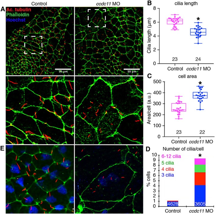
Mutations in CCDC11 (cfap53) have been identified in multiple patients with heterotaxy (Htx), a disorder of left-right (LR) patterning of the internal organs. In Xenopus, depletion of Ccdc11 causes defects in LR patterning, recapitulating the patient phenotype. Upon Ccdc11 depletion, monociliated cells of the Left-Right Organizer (LRO) exhibit multiple cilia per cell. Unexpectedly, we found that Ccdc11 is necessary for successful cytokinesis, explaining the multiciliation phenotype observed in Ccdc11-depleted cells. The small GTPase RhoA is critical for cytokinesis, and our Ccdc11 depletion phenotypes are reminiscent of RhoA loss of function. Here, we demonstrate that during cytokinesis CCDC11 is localized to the cytokinetic contractile ring overlapping with RhoA, and CCDC11 regulates total RhoA protein levels. Our results connect CCDC11 to cytokinesis and LR patterning via RhoA regulation, providing a potential mechanism for heterotaxy disease pathogenesis.

Keywords: Xenopus; CFAP53; RhoA; cilia; congenital heart disease; cytokinesis; left–right patterning.


Conflict of interest statement

The authors declare no conflicts of interest.

Figures

FIGURE 1
FIGURE 1
CCDC11 depletion alters cilia morphology and leads to multinucleation and multiciliation in Xenopus LRO cells. (A) Cilia in control and ccdc11 morphant (ccdc11 MO) Xenopus LROs marked by anti‐acetylated α‐tubulin (red) and F‐actin marked by phalloidin (green). Scale bar = 50 μm. (B) Length of cilia in uninjected control and ccdc11 morphant Xenopus LROs. Data is presented as box plot with 95% confidence interval. “n” = number of embryos. Results represent 3 independent experiments. (C) Average cell apical surface area of uninjected control and ccdc11 morphant Xenopus LROs. Data is presented as box plot with 95% confidence interval. “n” = number of embryos. Results represent 3 independent experiments. (D) Percent of cells with multiple cilia in uninjected controls and ccdc11 morphant Xenopus LROs. “n” = number of cells. Results represent 3 independent experiments. ★ = p < 0.001. (E) Xenopus left–right organizer (LRO) of a ccdc11 morphant embryo showing a cell with multiple nuclei and cilia compared to cells with one nucleus and one cilium in uninjected controls.
FIGURE 2
FIGURE 2
CCDC11 depletion leads to failed cytokinesis and multinucleation in Xenopus tropicalis. (A–D) Percentage of cells with multiple nuclei in (A, B) control MO, or depletion of Ccdc11 by (C, D) ccdc11 MO, or (E, F) ccdc11 CRISPR. Ccdc11 MO or CRISPR were injected in embryos at one cell stage. “n” = number of cells. Scale bar = 20 μm. Results represent 3 independent experiments. (G) Live imaging of Xenopus tropicalis embryo expressing utrophin calponin homology domain‐ (UtrCH‐) GFP at Nieuwkoop and Faber stage 10 (pre‐gastrulation) showing that cytokinesis is disrupted during actomyosin contractile ring ingression in ccdc11 morphant. The results represent 2 independent experiments.
FIGURE 3
FIGURE 3
CCDC11 depletion leads to failed cytokinesis and multinucleation in human U2OS cells. (A, B) Percentage of human U2OS cells with multiple nuclei in response to CCDC11 depletion by siRNA. “n” = number of cells. Scale bar = 50 μm. Results represent 3 independent experiments. (C) Western blot showing loss of CCDC11 protein in human U2OS cells transfected with CCDC11 siRNA. Results represent 2 independent experiments. (D) Western blot showing the antibody does not detect native Xenopus tropicalis Ccdc11 protein but can specifically detect human CCDC11 microinjected in Xenopus tropicalis embryos in a dose‐dependent manner. The results represent 2 independent experiments. ★ = p < 0.001.
FIGURE 4
FIGURE 4
Domains important for Ccdc11 localization to the contractile ring and midbody during cytokinesis. Live imaging of mNEON‐CCDC11 and fragments in blastomeres of Xenopus laevis embryos (Nieuwkoop and Faber stage 5–7). Time 0 is the time membrane ingression is first detectable. Arrowheads indicate the position of the contractile ring/midbody. Green arrowheads indicate strong accumulation of CCDC11 at the contractile ring, while white arrowheads indicate weak CCDC11 accumulation at the contractile ring. Yellow arrowheads indicate accumulation of CCDC11 at the midbody. Full length CCDC11 localizes to the cleavage furrow prior to ingression and the midbody following cytokinesis. D1 accumulates weakly at the contractile ring and strongly at the midbody. D2 accumulates weakly at the contractile ring. D3 accumulates strongly at the contractile ring. D1/D2 accumulates weakly at the contractile ring, indicating that D3 is necessary and sufficient for strong CR localization. D2/D3 accumulates strongly at the contractile ring and at the midbody, indicating that while D1 is sufficient for contractile ring localization, it is not required. Scale bar = 100 μm.
FIGURE 5
FIGURE 5
Ccdc11 co‐localizes with RhoA to the contractile ring and midbody in dividing U2OS cells and is important to maintain RhoA protein levels post‐transcriptionally. (A) Ccdc11 co‐localizes with RhoA in the contractile ring and midbody during cytokinesis in U2OS cells. Scale bars are 15 μm for Interphase and Early Anaphase and 10 μm for Late Anaphase and Cytokinesis. The results represent two independent experiments. (B–D) Ccdc11 depletion decreases RhoA accumulation at the cell junctions was measured using a line scan (white line) (B, C). RhoA accumulation at the contractile ring was measured, and the intensity at the ring was normalized with membrane expression (white box) (B, D) in the U2OS cells. The error bars represent the standard error. n = number of cells. The results represent three independent experiments. (E) Western blot showing a reduction in RhoA protein in human U2OS cells transfected with CCDC11 siRNA. The results represent three independent experiments. (F) Quantitative PCR data showing fold change mRNA levels of RhoA in CCDC11 siRNA compared to controls. The results represent three independent experiments. ★ = p < 0.001.

Similar articles

References

    1. Agarwal, R. , Varghese R., Jesudian V., and Moses J.. 2021. “The Heterotaxy Syndrome: Associated Congenital Heart Defects and Management.” Indian Journal of Thoracic and Cardiovascular Surgery 37: 67–81. - PMC - PubMed
    1. Ali, A. , Veeranki S. N., Chinchole A., and Tyagi S.. 2017. “MLL/WDR5 Complex Regulates Kif2A Localization to Ensure Chromosome Congression and Proper Spindle Assembly During Mitosis.” Developmental Cell 41, no. 605–622: e7. - PubMed
    1. Babu, D. , and Roy S.. 2013. “Left‐Right Asymmetry: Cilia Stir Up New Surprises in the Node.” Open Biology 3: 130052. - PMC - PubMed
    1. Bailey, J. K. , Fields A. T., Cheng K., et al. 2015. “WD Repeat‐Containing Protein 5 (WDR5) Localizes to the Midbody and Regulates Abscission.” Journal of Biological Chemistry 290: 8987–9001. - PMC - PubMed
    1. Basu, B. , Lake A. V. R., China B., et al. 2023. “Racgap1 Knockdown Results in Cells With Multiple Cilia Due to Cytokinesis Failure.” Annals of Human Genetics 88: 45–57. - PMC - PubMed

LinkOut - more resources