. 2024 Nov 19;98(11):e0094724.
doi: 10.1128/jvi.00947-24. Epub 2024 Oct 31.

Cyclophilin A facilitates HIV-1 integration

Affiliations

Cyclophilin A facilitates HIV-1 integration

Adrian Padron et al. J Virol. .

Abstract

Cyclophilin A (CypA) binds to the HIV-1 capsid to facilitate reverse transcription and nuclear entry and counter the antiviral activity of TRIM5α. Interestingly, recent studies suggest that the capsid enters the nucleus of an infected cell and uncoats prior to integration. We have previously reported that the capsid protein regulates HIV-1 integration. Therefore, we probed whether CypA-capsid interaction also regulates this post-nuclear entry step. First, we challenged CypA-expressing (CypA+/+) and CypA-depleted (CypA-/-) cells with HIV-1 and quantified the levels of provirus. CypA-depletion significantly reduced integration, an effect that was independent of CypA's effect on reverse transcription, nuclear entry, and the presence or absence of TRIM5α. In addition, cyclosporin A, an inhibitor that disrupts CypA-capsid binding, inhibited proviral integration in CypA+/+ cells but not in CypA-/- cells. HIV-1 capsid mutants (G89V and P90A) deficient in CypA binding were also blocked at the integration step in CypA+/+ cells but not in CypA-/- cells. Then, to understand the mechanism, we assessed the integration activity of the HIV-1 preintegration complexes (PICs) extracted from acutely infected cells. PICs from CypA-/- cells retained lower integration activity in vitro compared to those from CypA+/+ cells. PICs from cells depleted of both CypA and TRIM5α also had lower activity, suggesting that CypA's effect on PIC was independent of TRIM5α. Finally, CypA protein specifically stimulated PIC activity, as this effect was significantly blocked by CsA. Collectively, these results provide strong evidence that CypA directly promotes HIV-1 integration, a previously unknown role of this host factor in the nucleus of an infected cell.

Importance: Interaction between the HIV-1 capsid and host cellular factors is essential for infection. However, the molecular details and functional consequences of viral-host factor interactions during HIV-1 infection are not fully understood. Over 30 years ago, Cyclophilin A (CypA) was identified as the first host protein to bind to the HIV-1 capsid. Now it is established that CypA-capsid interaction promotes reverse transcription and nuclear entry of HIV-1. In addition, CypA blocks TRIM5α-mediated restriction of HIV-1. In this report, we show that CypA promotes the post-nuclear entry step of HIV-1 integration by binding to the viral capsid. Notably, we show that CypA stimulates the viral DNA integration activity of the HIV-1 preintegration complex. Collectively, our studies identify a novel role of CypA during the early steps of HIV-1 infection. This new knowledge is important because recent reports suggest that an operationally intact HIV-1 capsid enters the nucleus of an infected cell.

Keywords: Cyclophilin A (CypA); capsid; human immunodeficiency virus (HIV); integration; reverse transcription nuclear entry.


Conflict of interest statement

The authors declare no conflict of interest.

Figures
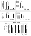
Fig 1
Fig 1
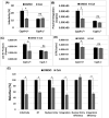
CypA depletion reduces HIV-1 integration. (A) Effects of CypA depletion on HIV-1 infectivity. Parental (CypA+/+) Jurkat T cells spinoculated with HIV-1.GFP reporter virus in the absence or presence of efavirenz and raltegravir. In parallel, the CypA-/- Jurkat T cells were spinoculated with HIV-1.GFP reporter virus. Cells were cultured for 24 hours and the intracellular GFP fluorescence (a marker of infectivity) was determined by FACS. (B) Effects of CypA depletion on HIV-1 integration. Integrated viral DNA (proviral DNA) was quantified by Alu-gag nested PCR assay of total DNA isolated from the infected cells. Copy numbers were calculated from standard curves generated in parallel using 10-fold serial dilutions of known copy numbers (100–108) of the HIV-1 proviral molecular clone during the second round qPCR. (C and D) Effects of CypA depletion on HIV-1 reverse transcription and nuclear entry. Late RT products (C) and 2-LTR circles (D) in the total DNA were quantified by qPCR. Copy numbers were calculated from standard curves generated in parallel using 10-fold serial dilutions of known copy numbers (100–108) of an HIV-1 proviral molecular clone (for late RT products) or the p2LTR plasmid (for 2-LTR circles). (E) Comparative analysis of the effects of CypA-depletion on HIV-1 reverse transcription, nuclear entry, and proviral DNA integration. To analyze the specific effects of CypA on HIV-1 integration, the data of CypA-depletion on infection (Panel A), proviral integration (Panel B), late RT products (Panel C), and 2-LTR circles (Panel D) were plotted as percentages relative to the parental (CypA+/+) cells. The effect of CypA-depletion on nuclear entry efficiency was determined by calculating the ratio of copy number of 2-LTR circles to the respective late RT products, whereas proviral integration efficiency was determined by calculating the ratio of copy number of integrated viral DNAs to the corresponding late RT products. The data shown are mean values from three independent experiments with error bars representing the standard error of the mean (SEM). The P-values (*) represent statistical significance (P < 0.05).
Fig 2
Fig 2
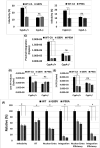
Reduced infectivity and proviral integration in CypA-depleted cells are sustained in cells lacking TRIM5α. (A) Effects of CypA and TRIM5α depletion on HIV-1 infectivity. Parental (CypA+/+), CypA-/-, and double knockout (CypA-/- & TRIM5α-/-) Jurkat T cells spinoculated with HIV-1.GFP reporter virus were cultured for 24 hours and the intracellular GFP fluorescence (a marker of infectivity) was measured by FACS. (B) Effects of CypA and TRIM5α depletion on HIV-1 integration. Proviral DNA was quantified by Alu-gag nested PCR assay of total DNA isolated from infected cells and copy numbers were calculated from standard curves as described in Fig. 1. (C and D) Effects of CypA and TRIM5α depletion on HIV-1 reverse transcription and nuclear entry. Late RT products (C) and 2-LTR circles (D) in the total DNA were quantified by qPCR and copy numbers were calculated from appropriate standard curves generated in parallel. The data shown are mean values from three independent experiments with error bars representing the SEM. The P-values (*) represent statistical significance (P < 0.05) and ns stands for a nonsignificant difference.
Fig 3
Fig 3
CsA markedly reduces HIV-1 integration. (A) Effects of CsA on HIV-1 infectivity. Parental (CypA+/+) and CypA-depleted (CypA-/-) Jurkat T cells inoculated with HIV-1.GFP virus in the presence of DMSO or 10 µM CsA were cultured for 24 hours and the intracellular GFP fluorescence (indicator of infectivity) was determined by FACS. (B) Effects of CsA on HIV-1 integration. Integrated viral DNA was quantified by Alu-gag nested PCR assay of total DNA isolated from infected cells and copy numbers were calculated from standard curves as described in Fig. 1. (C and D) Effects of CsA on HIV-1 reverse transcription and nuclear entry. Late RT products (C) and 2-LTR circles (D) in the total DNA from infected cells were determined by qPCR and copy numbers were calculated from standard curves generated in parallel. (E) Comparison of the effects of CsA on early steps of HIV-1 infection. To analyze the effects of CsA on HIV-1 integration, the data of CsA’s effect on virus infectivity (Panel A), proviral integration (Panel B), late RT products (Panel C), and nuclear entry (Panel D) were plotted as percentages relative to that of DMSO-treated cells. Nuclear entry efficiency and proviral integration efficiency were calculated as described in Fig. 1 legend. Data are mean values from at least three independent experiments with error bars representing the SEM. The P-values (*) represent statistical significance (P < 0.05) and ns stands for a nonsignificant difference.
Fig 4
Fig 4
HIV-1 CA mutant viruses with disrupted CypA binding are defective in proviral DNA integration. Effects of CA mutations (A) G89V and (B) P90A on HIV-1 infectivity. Parental (CypA+/+) and CypA-depleted (CypA-/-) Jurkat T cells inoculated with wild-type CA or mutant CA HIV-1 particles were cultured for 24 hours and intracellular GFP fluorescence (an indicator of infectivity) was determined by FACS. (C) Effects of CA mutations G89V and P90A on HIV-1 integration. Integrated viral DNA was quantified by Alu-gag nested PCR assay of total DNA from infected cells and copy numbers were calculated from standard curves as described in Fig. 1. (D and E) Effects of CA mutations G89V and P90A on HIV-1 reverse transcription and nuclear entry. Late RT products (D) and 2-LTR circles (E) in the total DNA from infected cells were quantified by qPCR and copy numbers were calculated from standard curves generated in parallel. (F) Comparison of the effects of CA mutations on early steps of HIV-1 infection. To analyze the specific effects of G89V or P90A on HIV-1 integration, the data of infectivity (A and B), proviral integration (C), late RT products (D), and nuclear entry (E) of the CA mutants were plotted as percentage relative to that of WT CA HIV-1 control. Nuclear entry efficiency and proviral integration efficiency were calculated as described in Fig. 1. Data are mean values from at least three independent experiments with error bars representing the SEM. The P-values (*) represent statistical significance (P < 0.05) and ns stands for a nonsignificant difference.
Fig 5
Fig 5
Effect of CypA-depletion on the integration activity of HIV-1 PICs. (A) PIC-associated integration activity measurements in vitro. (A) Parental (CypA+/+) Jurkat T cells were spinoculated with high titer HIV-1 particles (1,500 ng/mL of p24) and cultured for 5 hours followed by extraction of PIC-containing cytoplasmic extracts (Cy-PICs). In vitro assays using the Cy-PICs as the source of integration activity and quantification of viral DNA integration into an exogenous target DNA by nested PCR were carried out with appropriate controls. (B) Integration activity of PICs. In vitro integration assays of Cy-PICs extracted from CypA-expressing (CypA+/+) parental cells and from CypA-depleted (CypA-/-) Jurkat T cells were carried out and the copy numbers of integrated viral DNAs were determined. (C) Viral DNA content of the PICs. Copy numbers of viral DNA in the extracted PICs were quantified by qPCR and copy numbers were calculated from standard curves generated in parallel. (D) Specific PIC activity. The specific activity of the PIC-mediated viral DNA integration was determined by calculating the ratio of integrated viral DNA copy numbers (from panel B) to the corresponding PIC-associated viral DNA copy numbers (from panel C). Mean values from three independent experiments are shown with error bars representing the SEM. The P-values (*) represent statistical significance (P < 0.05).
Fig 6
Fig 6
CypA depletion-associated reduction in PIC integration activity is sustained in PICs from cells lacking TRIM5α as well. (A) Integration activity of PICs. Parental (CypA+/+), CypA-/-, and double knockout (CypA-/- & TRIM5α-/-) Jurkat T cells spinoculated with HIV-1 particles were cultured for 5 hours followed by extraction of Cy-PICs. In vitro integration assays of Cy-PICs were carried out as described in Materials and Methods and the copy numbers of integrated viral DNAs were determined by nested PCR as described in Fig. 5. (B) Viral DNA content of the PICs. Copy numbers of viral DNA in the extracted PICs were quantified by qPCR. (C) Specific PIC activity. The specific PIC activity was determined by calculating the ratio of integrated viral DNA copy numbers (panel B) to the corresponding PIC-associated viral DNA copy numbers (panel C) and plotted as percentages relative to the specific activity of Parental (CypA+/+) PICs. The data shown are mean values from three independent experiments with error bars representing the SEM. The P-values (*) represent statistical significance (P < 0.05).
Fig 7
Fig 7
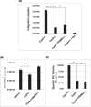
Effects of recombinant CypA protein in the absence or presence of CsA on PIC-associated integration activity in vitro. (A and B) Integration activity measurement in the presence of recombinant CypA protein. Integration activity in vitro of PICs extracted from CypA-expressing (CypA+/+) parental cells (A) and from CypA-depleted (CypA-/-) Jurkat T cells (B) were assessed in the absence (Control) or presence of varying concentrations (0–20 µM) of purified recombinant CypA protein, and the copy number of the integrated viral DNAs were determined by nested PCR. (C) Integration activity was plotted as fold change relative to the integration activity of the respective control PICs. (D) Integration activity measurement in the presence of CsA. PICs extracted from CypA-expressing (CypA+/+) cells were subjected to integration activity in the presence of CsA, CypA, or CsA +CypA. Mean values from three independent experiments are shown with error bars representing the SEM. The P-values (*) represent statistical significance (P < 0.05) and ns stands for nonsignificant.
Fig 8
Fig 8
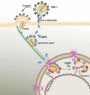
A hypothetical model depicting the mechanism by which CypA promotes HIV-1 DNA integration. The HIV-1 envelope spike binds to the CD4+ receptor and one of the co-receptors—CCR5 or CXCR4. The binding leads to the fusion of the viral and target cell membranes and the release of the capsid into the cytosol. HIV-1 capsid is transported along microtubules (shown as green elongated tube) where cytosolic CypA (deep blue colored structures) binds to the capsid shell en route to the nucleus. The operationally intact capsid containing the reverse transcribed viral DNA enters the nucleus through the NPC (magenta) and binds with other host factors such as CPSF6 as shown in deep red structures. Upon nuclear entry, CypA remains bound to the near intact capsid and then associated with viral replication complex (RTC/PIC) as shown in a light pink blob at the site of the integration. The capsid-bound CypA in the nucleus promotes the integration of the viral DNA into the host genomic DNA.

Update of

References

    1. van Schalkwyk C, Mahy M, Johnson LF, Imai-Eaton JW. 2024. Updated data and methods for the 2023 UNAIDS HIV estimates. J Acquir Immune Defic Syndr 95:e1–e4. doi:10.1097/QAI.0000000000003344 - DOI - PMC - PubMed
    1. Ostrowski MA, Chun TW, Justement SJ, Motola I, Spinelli MA, Adelsberger J, Ehler LA, Mizell SB, Hallahan CW, Fauci AS. 1999. Both memory and CD45RA+/CD62L+ naive CD4(+) T cells are infected in human immunodeficiency virus type 1-infected individuals. J Virol 73:6430–6435. doi:10.1128/JVI.73.8.6430-6435.1999 - DOI - PMC - PubMed
    1. Brenchley JM, Hill BJ, Ambrozak DR, Price DA, Guenaga FJ, Casazza JP, Kuruppu J, Yazdani J, Migueles SA, Connors M, Roederer M, Douek DC, Koup RA. 2004. T-cell subsets that harbor human immunodeficiency virus (HIV) in vivo: implications for HIV pathogenesis. J Virol 78:1160–1168. doi:10.1128/jvi.78.3.1160-1168.2004 - DOI - PMC - PubMed
    1. Le Douce V, Herbein G, Rohr O, Schwartz C. 2010. Molecular mechanisms of HIV-1 persistence in the monocyte-macrophage lineage. Retrovirology (Auckl) 7:32. doi:10.1186/1742-4690-7-32 - DOI - PMC - PubMed
    1. Sonza S, Mutimer HP, Oelrichs R, Jardine D, Harvey K, Dunne A, Purcell DF, Birch C, Crowe SM. 2001. Monocytes harbour replication-competent, non-latent HIV-1 in patients on highly active antiretroviral therapy. AIDS 15:17–22. doi:10.1097/00002030-200101050-00005 - DOI - PubMed

Publication types

MeSH terms

LinkOut - more resources