. 2024 Nov 19;98(11):e0109924.
doi: 10.1128/jvi.01099-24. Epub 2024 Oct 31.

HCMV strain- and cell type-specific alterations in membrane contact sites point to the convergent regulation of organelle remodeling

Affiliations

HCMV strain- and cell type-specific alterations in membrane contact sites point to the convergent regulation of organelle remodeling

William A Hofstadter et al. J Virol. .

Abstract

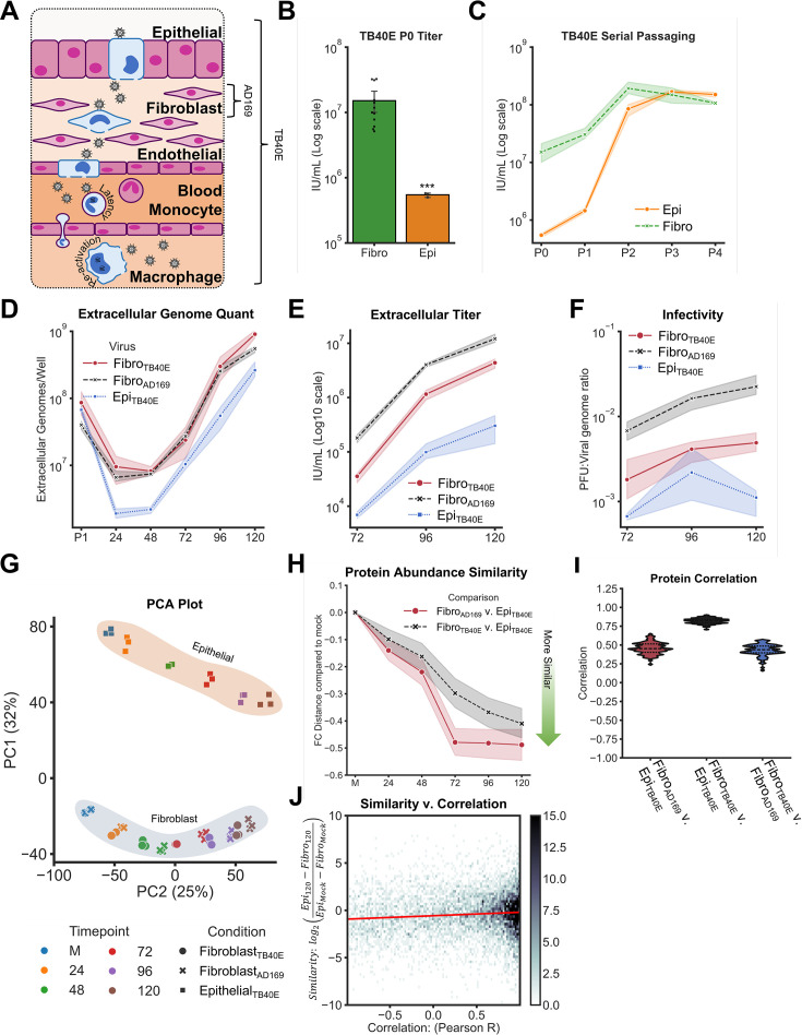
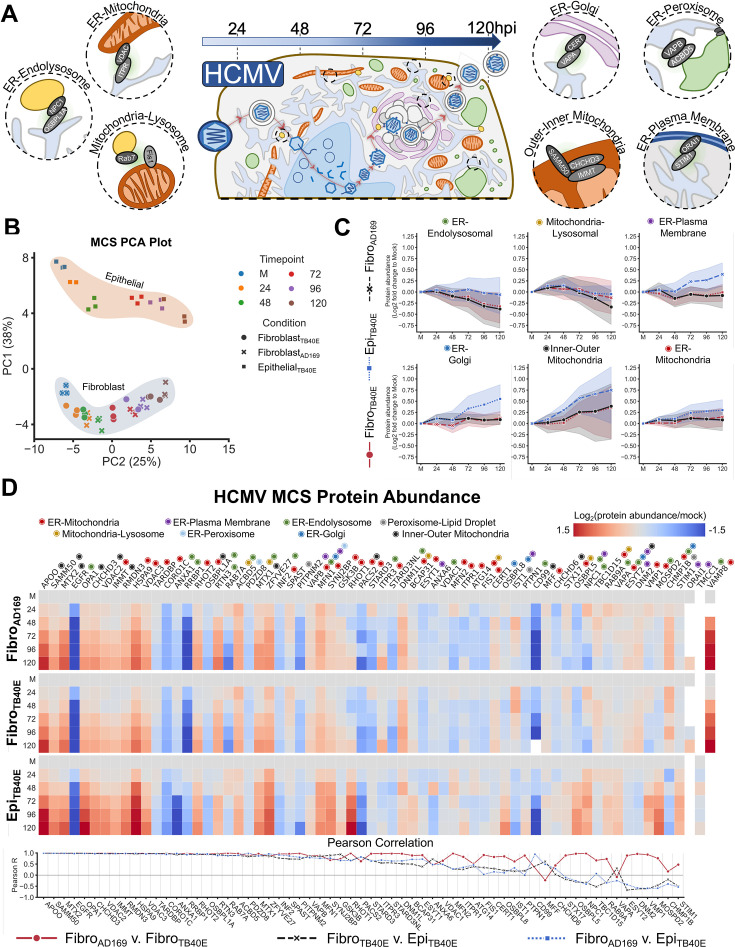
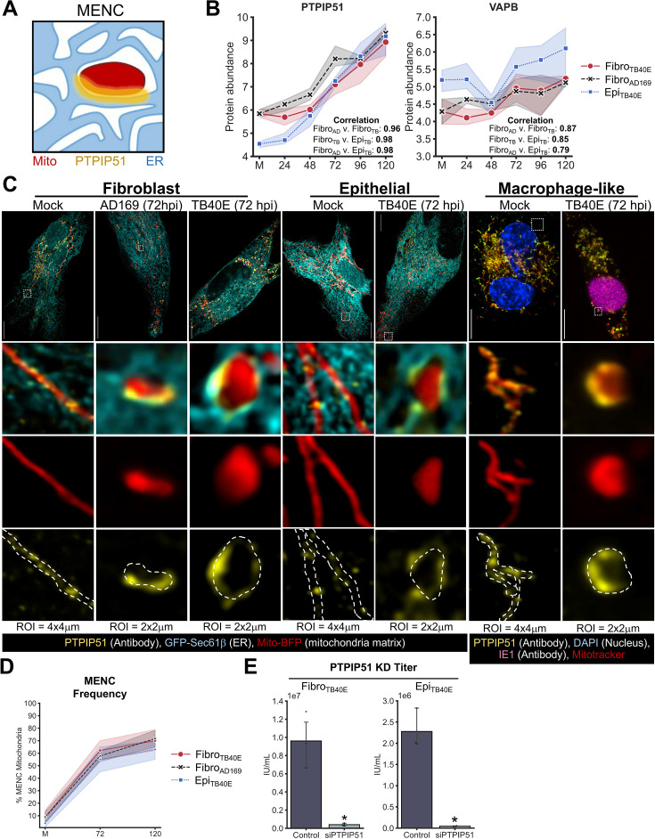
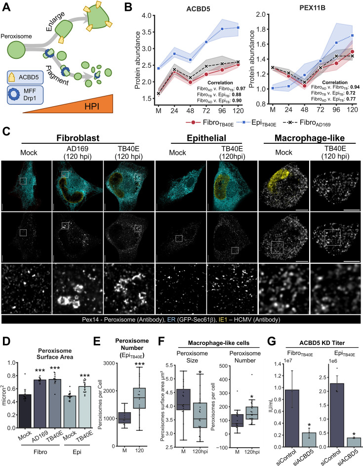
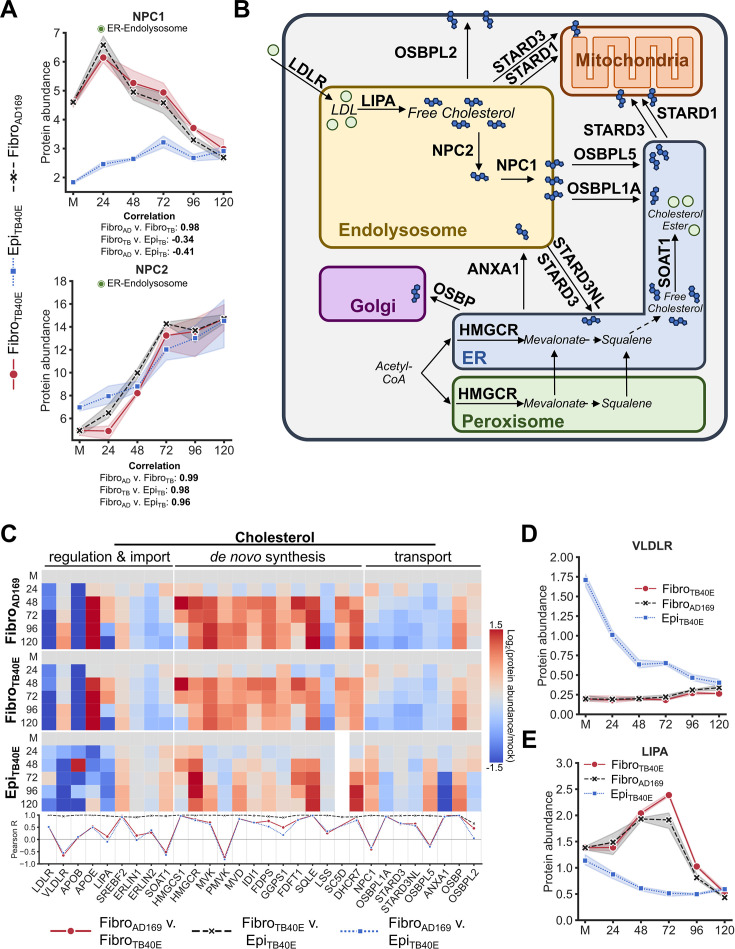
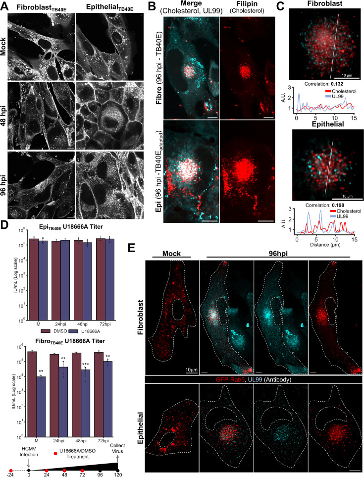
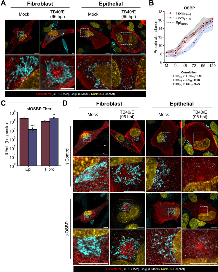
Viruses are ubiquitous entities that infect organisms across the kingdoms of life. While viruses can infect a range of cells, tissues, and organisms, this aspect is often not explored in cell culture analyses. There is limited information about which infection-induced changes are shared or distinct in different cellular environments. The prevalent pathogen human cytomegalovirus (HCMV) remodels the structure and function of subcellular organelles and their interconnected networks formed by membrane contact sites (MCSs). A large portion of this knowledge has been derived from fibroblasts infected with a lab-adapted HCMV strain. Here, we assess strain- and cell type-specific alterations in MCSs and organelle remodeling induced by HCMV. Integrating quantitative mass spectrometry, super-resolution microscopy, and molecular virology assays, we compare infections with lab-adapted and low-passage HCMV strains in fibroblast and epithelial cells. We determine that, despite baseline proteome disparities between uninfected fibroblast and epithelial cells, infection induces convergent changes and is remarkably similar. We show that hallmarks of HCMV infection in fibroblasts, mitochondria-endoplasmic reticulum (ER) encapsulations and peroxisome proliferation, are also conserved in infected epithelial and macrophage-like cells. Exploring cell type-specific differences, we demonstrate that fibroblasts rely on endosomal cholesterol transport while epithelial cells rely on cholesterol from the Golgi. Despite these mechanistic differences, infections in both cell types result in phenotypically similar cholesterol accumulation at the viral assembly complex. Our findings highlight the adaptability of HCMV, in that infections can be tailored to the initial cell state by inducing both shared and unique proteome alterations, ultimately promoting a unified pro-viral environment.IMPORTANCEHuman cytomegalovirus (HCMV) establishes infections in diverse cell types throughout the body and is connected to a litany of diseases associated with each of these tissues. However, it is still not fully understood how HCMV replication varies in distinct cell types. Here, we compare HCMV replication with lab-adapted and low-passage strains in two primary sites of infection, lung fibroblasts and retinal epithelial cells. We discover that, despite displaying disparate protein compositions prior to infection, these cell types undergo convergent alterations upon HCMV infection, reaching a more similar cellular state late in infection. We find that remodeling of the subcellular landscape is a pervasive feature of HCMV infection, through alterations to both organelle structure-function and the interconnected networks they form via membrane contact sites. Our findings show how HCMV infection in different cell types induces both shared and divergent changes to cellular processes, ultimately leading to a more unified state.

Keywords: HCMV; herpesvirus; membrane contact sites; organelle remodeling; proteomics.


Conflict of interest statement

The authors declare no conflict of interest.

Figures

Fig 1
Fig 1
Comparing HCMV infection kinetics and proteomic alterations in fibroblasts and epithelial cells. (A) Schematic of HCMV progression through many different cell types as might occur in a wild-type infection. (B) TB40/E replication titer in MRC5 fibroblasts (Fibro) and ARPE19 epithelial cells (Epi). (C) Serial passaging of TB40/E in ARPE19 epithelial cells increases the titer during infections in fibroblasts and epithelial cells. (D) Quantification of the number of released (extracellular) viral particles (genomes) during AD169 or TB40/E infection in epithelial cells and fibroblasts. (E) Quantification of the number of infectious released viral particles during AD169 or TB40/E infection in epithelial cells and fibroblasts. (F) Ratio of number of infectious virions to the number of total virions (genomes) released, as would indicate infectivity. (G) Principal component analysis (PCA) plot of proteomic data. (H) Change in protein abundance similarity between infections throughout HCMV infection. Similarity quantified as the Log2 fold change of the difference in protein abundance between infections at one timepoint divided by that difference in uninfected cells. (I) Pearson R correlation between infections for all proteins quantified. (J) Comparison between protein abundance trend correlation and protein abundance similarity at 120 h post infection for TB40/E infections in fibroblast and epithelial cells.
Fig 2
Fig 2
HCMV infection alters the MCS landscape independent of cell type. (A) Schematic of organelle remodeling across the HCMV replication cycle, as well as examples of MCSs. (B) PCA specifically of the MCS proteins quantified. (C) Average abundance trends for MCS proteins binned by what MCS(s) they localize to. (D) Top: quantification of MCS protein abundance trends across three different infections. Fisheyes mark what MCS(s) each protein localizes to. Blank squares indicate conditions where the protein was not detected. Bottom: Pearson R correlation between each infection for each MCS protein. Proteins are arranged in the same order as the above heatmap.
Fig 3
Fig 3
Mitochondria-ER encapsulations are hallmarks of HCMV infection, independent of cell type. (A) Schematic of a MENC, which are formed by ER tubules stably and asymmetrically cupping mitochondria. (B) Protein abundance trends for ER-mitochondria MCS proteins PTPIP51 (left) and VAPB (right) as quantified by DIA-MS. Pearson R correlations provided. (C) Representative images of MENC formation during HCMV infection in fibroblast, epithelial, and macrophage-like cells. Scale bars are 10 µm. (D) Quantification of percent mitochondria within MENCs in mock, 72, and 120 hpi cells. (E) HCMV titer following control (scramble) or PTPIP51-targeting siRNA treatment.
Fig 4
Fig 4
Peroxisome proliferation and enlargement are observed during HCMV infection in diverse cell types. (A) Schematic of peroxisome polarization during HCMV infection in fibroblasts, resulting in both proliferation and the formation of a subpopulation of enlarged and irregularly shaped peroxisomes. (B) Protein abundance trends for peroxisomal proteins ACBD5 (left) and PEX11β (right) as quantified by DIA-MS. Pearson R correlations provided. (C) Representative images of peroxisome dysregulation during HCMV infection in fibroblast, epithelial, and macrophage-like cells. Scale bars are 10 µm. Regions of interest (ROIs) are 10 × 10 µm. (D) Quantification of peroxisome surface area in uninfected cells as well as at 120 hpi for fibroblast and epithelial cell infections. (E) Quantification of peroxisome proliferation during HCMV infection in epithelial cells. (F) Quantification of peroxisome size (left) and proliferation (right) in THP-1 macrophage-like cells. (G) HCMV titer following control (scramble) or ACBD5-targeting siRNA treatment.
Fig 5
Fig 5
Proteomic comparison of cholesterol regulation during HCMV infection in different cell types. (A) Protein abundance trends for ER-endosomal MCS proteins NPC1 (upper) and NPC2 (lower) as quantified by DIA-MS. Pearson R correlations provided. (B) Schematic of some of the ways in which cholesterol is imported, synthesized, and transported throughout a cell. (C) Upper: quantification of cholesterol regulatory proteins. Lower: Pearson R correlation between each infection for each protein. Proteins are arranged in the same order as the above heatmap. (D and E) Protein abundance trends for ER-endosomal MCS proteins Very low-density lipoprotein receptor (VLDLR) (D) and LIPA (E) as quantified by DIA-MS.
Fig 6
Fig 6
Cholesterol accumulates in the HCMV assembly complex in different cell types via distinct mechanisms. (A) Representative images of cholesterol (Filipin III, white) distribution and accumulation across TB40/E HCMV infection in fibroblasts and epithelial cells. Scale bars are 10 µm. (B) Representative microscopy of cholesterol (Filipin III) accumulation in the HCMV assembly complex, marked by the viral protein UL99. Scale bars are 10 µm. (C) Regions of interest (ROIs) from Fig. 6B of the assembly complex. White lines indicate where the line scan was performed. Scale bar is 10 µm. (D) HCMV titers following treatment with either U18666A or control dimethyl sulfoxide (DMSO) at the timepoint indicated on the x-axis in either epithelial cells (upper) or fibroblasts (lower). At the bottom shows a schematic of the drug treatment timing. (E) Representative images of Rab5 endosome (red) redistribution to the assembly complex (UL99, cyan) during HCMV infection in fibroblast and epithelial cells.
Fig 7
Fig 7
Cholesterol transport to the Golgi is pro-viral during HCMV infection in epithelial cells but antiviral in fibroblasts. (A) Representative images of the Golgi reorganization during TB40/E HCMV infection. Images are max projections. All scale bars are 10 µm. (B) Protein abundance trend for OSBP as measured by DIA-MS. (C) HCMV titer following treatment with a control siRNA or a mix of two siRNAs targeting OSBP. (D) Representative images of cholesterol and Golgi reorganization following control or OSBP KD and TB40/E HCMV infection. Images are max projections. All scale bars are 10 µm.

Similar articles

Cited by

References

    1. Zamarin D, García-Sastre A, Xiao X, Wang R, Palese P. 2005. Influenza virus PB1-F2 protein induces cell death through mitochondrial ANT3 and VDAC1. PLoS Pathog 1:e4. doi:10.1371/journal.ppat.0010004 - DOI - PMC - PubMed
    1. Yoshizumi T, Ichinohe T, Sasaki O, Otera H, Kawabata S, Mihara K, Koshiba T. 2014. Influenza A virus protein PB1-F2 translocates into mitochondria via Tom40 channels and impairs innate immunity. Nat Commun 5:6–10. doi:10.1038/ncomms5713 - DOI - PubMed
    1. Gibbs JS, Malide D, Hornung F, Bennink JR, Yewdell JW. 2003. The influenza A virus PB1-F2 protein targets the inner mitochondrial membrane via a predicted basic amphipathic helix that disrupts mitochondrial function. J Virol 77:7214–7224. doi:10.1128/jvi.77.13.7214-7224.2003 - DOI - PMC - PubMed
    1. Pila-Castellanos I, Molino D, McKellar J, Lines L, Da Graca J, Tauziet M, Chanteloup L, Mikaelian I, Meyniel-Schicklin L, Codogno P, Vonderscher J, Delevoye C, Moncorgé O, Meldrum E, Goujon C, Morel E, de Chassey B. 2021. Correction: Mitochondrial morphodynamics alteration induced by influenza virus infection as a new antiviral strategy. PLoS Pathog 17:e1009485. doi:10.1371/journal.ppat.1009485 - DOI - PMC - PubMed
    1. Gerna G, Kabanova A, Lilleri D. 2019. Human cytomegalovirus cell tropism and host cell receptors. Vaccines (Basel) 7:70. doi:10.3390/vaccines7030070 - DOI - PMC - PubMed