. 2024 Oct 31;135(1):e182550.
doi: 10.1172/JCI182550.

Leptin signaling maintains autonomic stability during severe influenza infection in mice

Affiliations

Leptin signaling maintains autonomic stability during severe influenza infection in mice

Andrés R Muñoz-Rojas et al. J Clin Invest. .
No abstract available

Keywords: Endocrinology; Infectious disease; Influenza; Leptin.


Figures
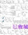
Figure 1
Figure 1. Leptin supplementation maintains autonomic stability during severe influenza infection.
(A) Kaplan-Meier curve of influenza A–infected WT lean and DIO mice and ob/ob mice and (B) influenza A–infected ob/ob mice supplemented with leptin. (CE) Serum levels of (C) IFN-β (D) CCL5, and (E) IL-12. (F and G) Whole-tissue RNA-Seq of (F) lung and (G) spleen. Average normalized expression values for the chosen gene clusters across time (left), and functional enrichment analysis for each cluster (right). (H) Weight, (I) number of immunocytes, and (J) proportion of cell types in spleens 8 days after infection. (K) Representative flow cytometry plots (left) and quantification (right) of Th1 cells in spleens. (L and M) Immunophenotyping of lungs 8 days after infection, showing the fold change in (L) proportions and (M) numbers of different cell types. (N) H&E staining of lungs. Normal, uninfected tissue is shown (left). Seven days after infection, both Ob-ctrl–infected (middle) and ob-leptin–infected (right) tissues showed attenuation of the epithelium (black double-headed arrows), necrotic epithelial cells (black arrows), peribronchiolar infiltrates of immune cells (white double-headed arrows), and luminal debris (black asterisk). Flu, influenza. Original magnification, ×20. Scale bars: 40 μm. (O and P) Viral titers of (O) lung homogenates (P) and serum. (Q) Oxygen saturation (R) heart rate, and (S) core temperature of ob/ob mice 7 days after infection. OB, ob/ob; DPI, days post infection; Mθ, macrophages; Alv Mθ, alveolar macrophages; Inter Mθ, interstitial macrophages; Trans Mθ, transitional macrophages; TCID50, 50% tissue culture infectious dose; bpm, beats per minute. *P < .05, **P < .01, ***P < .005, ****P < .001, by 2-tailed Student’s t test. Data represent the mean ± SEM.

References

    1. Milner JJ, et al. Obesity increases mortality and modulates the lung metabolome during pandemic H1N1 influenza virus infection in mice. J Immunol. 2015;194(10):4846–4859. doi: 10.4049/jimmunol.1402295. - DOI - PMC - PubMed
    1. Bell BB, et al. Differential contribution of POMC and AgRP neurons to the regulation of regional autonomic nerve activity by leptin. Mol Metab. 2018;8:1–12. doi: 10.1016/j.molmet.2017.12.006. - DOI - PMC - PubMed