. 2024 Oct 31;19(10):e0306676.
doi: 10.1371/journal.pone.0306676. eCollection 2024.

The evolutionarily conserved PhLP3 is essential for sperm development in Drosophila melanogaster

Affiliations

The evolutionarily conserved PhLP3 is essential for sperm development in Drosophila melanogaster

Christopher Petit et al. PLoS One. .

Abstract

Phosducin-like proteins (PhLP) are thioredoxin domain-containing proteins that are highly conserved across unicellular and multicellular organisms. PhLP family proteins are hypothesized to function as co-chaperones in the folding of cytoskeletal proteins. Here, we present the initial molecular, biochemical, and functional characterization of CG4511 as Drosophila melanogaster PhLP3. We cloned the gene into a bacterial expression vector and produced enzymatically active recombinant PhLP3, which showed similar kinetics to previously characterized orthologues. A fly strain homozygous for a P-element insertion in the 5' UTR of the PhLP3 gene exhibited significant downregulation of PhLP3 expression. We found these male flies to be sterile. Microscopic analysis revealed altered testes morphology and impairment of spermiogenesis, leading to a lack of mature sperm. Among the most significant observations was the lack of actin cones during sperm maturation. Excision of the P-element insertion in PhLP3 restored male fertility, spermiogenesis, and seminal vesicle size. Given the high level of conservation of PhLP3, our data suggests PhLP3 may be an important regulator of sperm development across species.


Conflict of interest statement

The authors have declared that no competing interests exist.

Figures

Fig 1
Fig 1. Sequence and structure conservation of PhLP3 proteins.
(A) Clustal Omega alignment of published sequences. Similar amino acids are underlaid in gray. Secondary structure predictions by JPred4 [11] are shown above the sequences with alpha helices in red and beta strands in yellow. The blue box indicates the thioredoxin domain. The conserved putative redox-active cysteine is highlighted in yellow. The inset table shows sequence identities between Drosophila melanogaster PhLP3 and the sequences in the alignment. Abbreviations and NCBI references: At = Arabidopsis thaliana (AEE78732; [12]), Pb = Plasmodium berghei (SCO61590; [4]), Ce = Caenorhabditis elegans (NP_498410; [2]), Dm = Drosophila melanogaster (AAL28410; this paper), Dr = Danio rerio (AAI65424; [13]), Hs = Homo sapiens (NP_005774; [4, 6]), Rn = Rattus norwegicus (NP_742051; [14]). (B) Phylogenetic tree of the aligned sequences. Alignment and phylogenetic tree were generated using EMBL-EBI Clustal Omega tools [15]. The scale represents the "length" of the branches, which indicates the evolutionary distance between the sequences in units of amino acid substitutions per site. Longer branches represent larger numbers of genetic changes.
Fig 2
Fig 2. Drosophila melanogaster sperm development.
(A) Stages of D. melanogaster spermatogenesis. Germline stem cells (GSCs; light blue) and cyst stem cells (CySCs; light green) are regulated and maintained by hub cells (magenta) at the apical tip. GSCs and CySCs divide asymmetrically to give rise to differentiating daughter cells, known as gonialblasts (dark blue) and somatic cyst cells (dark green), respectively. Gonialblasts undergo four mitotic divisions with incomplete cytokinesis, yielding cysts of 16 interconnected spermatogonia. Following mitosis, spermatogonia mature into spermatocytes, which grow and proceed through meiosis with incomplete cytokinesis to form 64 interconnected, haploid, round spermatids encased within a syncytial cyst. The post-meiotic maturation of spermatids is referred to as spermiogenesis. During this stage, spermatids extend axonemes, elongate nuclei, and are individualized by actin-based individualization complexes, also known as actin cones or investment cones (yellow triangles). Mature sperm coil and move into the seminal vesicle [24]. (B) Stages of nuclear elongation during spermiogenesis. Following meiosis, spermatid nuclei are round and begin to elongate, passing through the leaf and canoe stage to the final needle-like stage [25]. N: nucleus, BB: basal body, dc: dense complex, ca: centriolar adjunct, and Ax: axoneme (axoneme not to scale).
Fig 3
Fig 3. Organization of the PhLP3 gene.
The gene is located on chromosome 3 in the D. melanogaster genome (10854501–10856412) (FlyBase: FB2023_06, released December 12, 2023). The transcription start sites (TSS) are indicated. Intron 1 is located in the 5’ UTR. Exon 2 contains a splice site resulting in two transcripts, RA and RB, containing exon 2A and exon 2B, respectively. The transcripts differ only in their 5’ UTR regions and translate into identical proteins. Green boxes indicate coding regions of exons, while light blue boxes indicate untranslated regions. Protein domains are indicated according to secondary structure prediction JPred [11] as well as structural alignment of human PhLP2A using SWISS-MODEL [34]. Primers used for RT-qPCR are indicated in purple and pink.
Fig 4
Fig 4. AlphaFold and hypothetical model of Drosophila melanogaster PhLP3.
(A) AlphaFold prediction of the complete D. melanogaster PhLP3 structure (Q9VGV8; [29, 30]) showing the characteristic PhLP organization. Amino acid stretches missing at the N- and C-termini in the SWISS-MODEL are indicated. (B) Structure superposition of D. melanogaster PhLP3 (DmPhLP3) with human PhLP2A (hPhLP2A; PDB 7nvm: entity 11; [32]). Blue represents hPhLP2A, while red and green structures represent DmPhLP3. The green portions indicate unstructured segments, including loops and turns. The enhancement box shows the superposition for the conserved putative redox-active cysteines CYS95 (DmPhLP3) and CYS127 (hPhLP2A). Structural alignment and rendering were prepared using PyMOL (Pymol.org, The PyMOL Molecular Graphics System, Version 1.2r3pre, Schrödinger, LLC). Alpha helix a2’ in the SWISS-MODEL is part of helix a2 in the AlphaFold model. (C) Sequence alignment of DmPhLP3 with hPhLP2A.The pairwise sequence alignment was generated using Clustal Omega [15]. Amino acids are colored by polarity. The open green box represents the thioredoxin domain, and the blue bar above the sequence indicates the sequence portion modeled by SWISS-MODEL shown in B. * = identical AA;: = highly similar AA;. = similar AA. NCBI reference sequences: hPhLP2A NP_076970 [10], DmPhLP3 AAL28410.
Fig 5
Fig 5. Purification and enzyme activity of PhLP3.
(A) SDS gel following protein purification of recombinant His-tagged PhLP3. Washes (W) and elutions with increasing imidazole concentrations are indicated. (B) Principal setup of an in vitro thioredoxin reduction assay. The oxidation of NADPH to NADP is monitored at 340 nm. Red arrows indicate electron-flow from NADPH to Thioredoxin reductase (TrxR) to thioredoxin (Trx-1) and then to PhLP3, which will be reduced in the process. (C) Michaelis Menten diagram showing activity of PhLP3 with increasing thioredoxin concentrations. The indicated Km and Vmax values were determined using curve fitting. The values were verified by generating a Lineweaver Burk plot (inset).
Fig 6
Fig 6. Expression of PhLP3 in the testes.
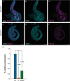
(A-F) Fluorescent in situ hybridization for PhLP3. PhLP3 probe (magenta) and anti-Vasa to label germ cells (cyan). Scale bar is 100 μm. (A) PhLP3+/+ testes exhibit expression in germ cells from spermatogonia through early spermatid stages (n = 59). (B) Germ cells immunostained with anti-Vasa (cyan). (C) In situ hybridization for PhLP3 (magenta). (D) PhLP3-/- testes exhibit a loss of PhLP3 expression throughout the testis (n = 36). (E) Germ cells immunostained with anti-Vasa (cyan). (F) In situ hybridization for PhLP3 (magenta). (G) RT-qPCR shows downregulation of PhLP3 expression in testes from males homozygous for the P-element insertion in the 5’ UTR of PhLP3 (-/-), compared to wild-type (+/+) levels. Excision of the P-element (ΔP/ΔP) partially rescues PhLP3 expression.
Fig 7
Fig 7. Fertility tests.
Control males (w1118), PhLP3-/- (PhLP3EY13373/EY13373) males or P element excision males (PhLP3ΔP/ΔP) were mated to w1118 virgin females to assess fertility. The average number of progeny per cross was counted. Individual measurements for each cross are shown with the average and standard deviation indicated. *** indicates p<0.001 and ** indicates p<0.01 based on pairwise analysis by Welch’s t-test; ns = not significant.
Fig 8
Fig 8. Mutation of PhLP3 causes arrest of spermiogenesis.
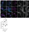
(A-C) Control PhLP3+/+ testes have DAPI clusters and actin-based individualization complexes (n = 40). Nuclei stained with DAPI (cyan) and actin-based individualization complexes stained with phalloidin (magenta). (B-C) Higher magnification view of testis in (A). Arrows indicate individualization complexes. (D-E) Control PhLP3+/+ seminal vesicle. (D) Seminal vesicle is large with mature sperm (n = 21). Nuclei stained with DAPI. (E) Zoomed in view of the seminal vesicle from (D) filled with mature sperm. (F-H) PhLP3-/- testes have dispersed DAPI clusters, less elongated nuclei, and no actin-based individualization complexes (n = 38). Nuclei stained with DAPI (cyan) and actin-based individualization complexes stained with phalloidin (magenta). (G-H) Higher magnification view of testis in (F). (I-J) PhLP3-/- seminal vesicle. (I) Seminal vesicle is smaller than control and lacks sperm (n = 15). Nuclei stained with DAPI. (J) Zoomed in view of the seminal vesicle from (I) lacking mature sperm. (K-M) Excision of the P element in PhLP3 (PhLP3ΔP/ΔP) rescues the mutant phenotype. Testis contain DAPI clusters and individualization complexes (n = 23). Nuclei stained with DAPI (cyan) and actin-based individualization complexes stained with phalloidin (magenta). (L-M) Higher magnification view of testis in (K). Arrows indicate DAPI clusters with individualization complexes. (N-O) PhLP3ΔP/ΔP seminal vesicle. (N) Seminal vesicle contains mature sperm (n = 19). Nuclei stained with DAPI. (O) Zoomed in view of the seminal vesicle from (N) filled with mature sperm. (P) Cross section area of the seminal vesicle (n = 10 for each genotype). Individual measurements are shown with the average and standard deviation indicated. **** indicates p<0.0001 and *** indicates p<0.001 based on ordinary one-way ANOVA; ns = not significant. Scale bars are 100 μM in A-D, F-I, and K-N. Scale bar is 50 μM in E, J, and O.
Fig 9
Fig 9. Morphology of spermatids in PhLP3 mutants.
(A-C) Control PhLP3+/+ testes have clusters of spermatid nuclei undergoing multiple stages of spermiogenesis, as indicated by DAPI (cyan). Individualizing spermatids are stained with phalloidin (magenta). (n = 40). (B) PhLP3-/- testes have dispersed spermatid nuclei at various stages of elongation but lack needle-like nuclei, as indicated by DAPI staining (cyan). Individualization complexes are not observed by phalloidin staining (magenta) (n = 37). (A, D) Arrowheads label canoe stage nuclei. Dashed arrows label leaf stage nuclei. Solid arrows label needle-like nuclei. Scale bars are 10 μM. Images are maximum intensity Z projections of whole-mount testes.
Fig 10
Fig 10. Effects of PhLP3 RNAi on mature sperm production.
(A) RT-qPCR demonstrates a 77% reduction in PhLP3 expression by RNA-interference (RNAi) in comparison to that of control (bam-Gal4) samples. (B) Fertility cross data showing the number of progeny resulting from four virgin female w1118 flies crossed with three male bam-Gal4 control flies or three male bam-Gal4>PhLP3-RNAi flies. Individual measurements for each cross are shown with the average and standard deviation indicated. **** indicates p<0.0001 based on Welch’s t-test. (C-F) bam-Gal4 control testis (n = 47). Whole testis stained with DAPI (cyan) stain to visualize nuclei and phalloidin (magenta) to visualize actin-based individualization complexes. (D-F) Higher magnification image of testis from (C). (D) Arrows indicate colocalized DAPI clusters with actin-based individualization complexes (magenta). (E) Arrows indicate actin-based individualization complexes. (F) Arrows indicate clusters of aligned, elongating spermatid nuclei stained with DAPI. (G) bam-Gal4 control seminal vesicle filled with mature sperm indicated by bracket (n = 24). (H) Zoomed in view of seminal vesicle from (G) filled with sperm (G). (I-L) bam-Gal4>PhLP3-RNAi testis (n = 29). Whole testis stained with DAPI (cyan) stain to visualize nuclei and phalloidin (magenta) to visualize actin-based individualization complexes. (J-L) Higher magnification image of testis from (I). (J) There is little colocalization of DAPI with actin-based individualization complexes. (K) Arrows indicate scattered actin cones. (L) Arrows indicate scattered, elongating spermatid nuclei that fail to cluster as compared to controls. (M) bam-Gal4>PhLP3-RNAi seminal vesicle indicated by bracket is reduced in size and lacks mature sperm (n = 19). (N) Zoomed in view of the seminal vesicle from (M) lacking mature sperm. Scale bar is 100 μM in C-G and I-M. Scale bar is 50 μM in H and N.
Fig 11
Fig 11. Model of defects observed in PhLP3 mutants.
Spermatid nuclei are misshapen and scattered, failing to reach the needle-like stage. Individualization complexes are not observed in PhLP3 mutants and sperm do not transfer to the seminal vesicle, leading to reduced seminal vesicle size.

References

    1. Gaudet R, Bohm A, Sigler PB. Crystal structure at 2.4 angstroms resolution of the complex of transducin betagamma and its regulator, phosducin. Cell. 1996;87: 577–588. doi: 10.1016/s0092-8674(00)81376-8 - DOI - PubMed
    1. Ogawa S, Matsubayashi Y, Nishida E. An evolutionarily conserved gene required for proper microtubule architecture in Caenorhabditis elegans. Genes Cells Devoted Mol Cell Mech. 2004;9: 83–93. doi: 10.1111/j.1356-9597.2004.00708.x - DOI - PubMed
    1. Putonti C, Quach B, Kooistra RL, Kanzok SM. The evolution and putative function of phosducin-like proteins in the malaria parasite Plasmodium. Infect Genet Evol J Mol Epidemiol Evol Genet Infect Dis. 2013;13: 49–55. doi: 10.1016/j.meegid.2012.08.023 - DOI - PubMed
    1. Kooistra RL, David R, Ruiz AC, Powers SW, Haselton KJ, Kiernan K, et al.. Characterization of a protozoan Phosducin-like protein-3 (PhLP-3) reveals conserved redox activity. PloS One. 2018;13: e0209699. doi: 10.1371/journal.pone.0209699 - DOI - PMC - PubMed
    1. Lukov GL, Hu T, McLaughlin JN, Hamm HE, Willardson BM. Phosducin-like protein acts as a molecular chaperone for G protein betagamma dimer assembly. EMBO J. 2005;24: 1965–1975. doi: 10.1038/sj.emboj.7600673 - DOI - PMC - PubMed

Substances

LinkOut - more resources