. 2024 Oct 31;20(10):e1012683.
doi: 10.1371/journal.ppat.1012683. eCollection 2024 Oct.

B cell receptor dependent enhancement of dengue virus infection

Affiliations

B cell receptor dependent enhancement of dengue virus infection

Chad Gebo et al. PLoS Pathog. .

Abstract

Dengue virus (DENV) is the causative agent of dengue, a mosquito-borne disease that represents a significant and growing public health burden around the world. A unique pathophysiological feature of dengue is immune-mediated enhancement, wherein preexisting immunity elicited by a primary infection can enhance the severity of a subsequent infection by a heterologous DENV serotype. A leading mechanistic explanation for this phenomenon is antibody dependent enhancement (ADE), where sub-neutralizing concentrations of DENV-specific IgG antibodies facilitate entry of DENV into FcγR expressing cells such as monocytes, macrophages, and dendritic cells. Accordingly, this model posits that phagocytic mononuclear cells are the primary reservoir of DENV. However, analysis of samples from individuals experiencing acute DENV infection reveals that B cells are the largest reservoir of infected circulating cells, representing a disconnect in our understanding of immune-mediated DENV tropism. In this study, we demonstrate that the expression of a DENV-specific B cell receptor (BCR) renders cells highly susceptible to DENV infection, with the infection-enhancing activity of the membrane-restricted BCR correlating with the ADE potential of the IgG version of the antibody. In addition, we observed that the frequency of DENV-infectible B cells increases in previously flavivirus-naïve volunteers after a primary DENV infection. These findings suggest that BCR-dependent infection of B cells is a novel mechanism immune-mediated enhancement of DENV-infection.


Conflict of interest statement

The authors have declared that no competing interests exist.

Figures

Fig 1
Fig 1. Expression of DENV-specific BCRs renders cells susceptible to DENV-infection.
A) Schematic representation of DENV/IgG immune complex binding to an FcgR (left), and a DENV virion binding to a DENV-specific BCR (right). Created in BioRender. B) Expression and DENV E protein binding activity of 293T cells transfected with the indicated tmIgG constructs. C) DENV-1 RVP infection of 293T cells transfected with the indicated tmIgG constructs. DC-SIGN and tmIgG conditions gated on receptor-positive cells. D) Quantification of DENV-1 RVP infection of 293T cells transfected with DC-SIGN or the indicated tmIgG constructs. Error bars +/- SEM. Experiments were conducted across three biological replicates in triplicate. E) ADE activity of the indicated DENV-specific IgG mAbs at a 2.5ug/ml concentration in K562 cells utilizing a DENV-1 RVP. F) Titration of ADE activity of the indicated DENV-specific IgG mAbs in K562 cells utilizing a DENV-1 RVP. G) Plot of the ADE and BDE results described above. Error bars +/- SEM.
Fig 2
Fig 2. DENV-specific B cells are susceptible to ex vivo DENV infection.
A) Whole virus binding profiles of IgG antibody (1 μg) from cross-reactive 7B9 and ZIKV-specific 2F3 mAb to DENV1-4 and ZIKV by ELISA. B) Surface IgG surface expression of immortalized 7B9 and C) 2F3 B cells. D) Quantification of DENV-2 infection of immortalized 7B9 (DENVreact) and 2F3 (ZIKVreact) B cells at MOI of 10. Error bars +/- SEM. Experiments were conducted across two biological replicates in triplicate E) Pie charts of DENV-2 infected 7B9 B cell proportions in control and DENV-2 inoculated conditions as assessed by scRNAseq. F) Correlation plot of DENV (+) and DENV (-) sense RNA expression in DENV-2 exposed 7B9 B cells as assessed by scRNAseq. G) Representative flow cytometry plot showing the frequency of DENV-4 infected CD19+ B cells under the indicated culture conditions. Detection of DENV-infected cells was performed by staining fixed/permeabilized cells with a FITC-conjugated 4G2 antibody. H) Quantification of DENV-4 infected CD19+ B cells under the indicated culture conditions. Error bars +/- SEM. **** p < 0.001, One-way ANOVA with Dunnett’s multiple comparisons test, with a single pooled variance. Experiments were conducted across three biological replicates in triplicate.
Fig 3
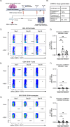
Fig 3. Frequency of DENV-infectible B cells increases following primary DENV Infection.
A) Schematic representation of the dengue human infection model and in vitro DENV-1 RVP infection assay. Created in BioRender. B) Key DHIM study characteristic and performance parameters. C) Representative flow cytometry plots showing the frequency of DENV-1 RVP infectible viable B cells (CD3-CD19+) in PBMC samples obtained 0-, 28-, and 90-days post DENV-3 infection. D) Quantification of DENV-1 RVP infectible B cells from all subjects included in this analysis. E) Representative flow cytometry plots showing the frequency of DENV-1 RVP infectible viable T cells (CD3+CD19-) in PBMC samples obtained 0-, 28-, and 90-days post DENV-3 infection. F) Quantification of DENV-1 RVP infectible T cells from all subjects included in this analysis. G) Representative flow cytometry plots showing the frequency of DENV-1 RVP infectible viable monocytes (CD3-CD19-CD14+) in PBMC samples obtained 0-, 28-, and 90-days post DENV-3 infection. H) Quantification of DENV-1 RVP infectible monocytes from all subjects included in this analysis. Error bars +/- SEM. ** p < 0.01, paired one-way ANOVA with correction for multiple comparisons (Friedman test with Dunn’s multiple comparisons test).

References

    1. Organization WH. Dengue and severe dengue World Health Organization [updated March 17, 2023; cited 2024 April 7]. Available from: https://www.who.int/news-room/fact-sheets/detail/dengue-and-severe-dengue.
    1. Tantracheewathorn T, Tantracheewathorn S. Risk factors of dengue shock syndrome in children. J Med Assoc Thai. 2007;90(2):272–7. . - PubMed
    1. Thein S, Aung MM, Shwe TN, Aye M, Zaw A, Aye K, et al.. Risk factors in dengue shock syndrome. Am J Trop Med Hyg. 1997;56(5):566–72. doi: 10.4269/ajtmh.1997.56.566 . - DOI - PubMed
    1. Bhatt S, Gething PW, Brady OJ, Messina JP, Farlow AW, Moyes CL, et al.. The global distribution and burden of dengue. Nature. 2013;496(7446):504–7. Epub 2013/04/09. doi: 10.1038/nature12060 ; PubMed Central PMCID: PMC3651993. - DOI - PMC - PubMed
    1. Salje H, Cummings DAT, Rodriguez-Barraquer I, Katzelnick LC, Lessler J, Klungthong C, et al.. Reconstruction of antibody dynamics and infection histories to evaluate dengue risk. Nature. 2018;557(7707):719–23. Epub 2018/05/26. doi: 10.1038/s41586-018-0157-4 ; PubMed Central PMCID: PMC6064976. - DOI - PMC - PubMed

MeSH terms