. 2024 Nov;386(6721):516-525.
doi: 10.1126/science.adp8721. Epub 2024 Oct 31.

Brain malformations and seizures by impaired chaperonin function of TRiC

Florian Kraft #  1 Piere Rodriguez-Aliaga #  2 Weimin Yuan #  3 Lena Franken  1 Kamil Zajt  4 Dimah Hasan  5 Ting-Ting Lee  2 Elisabetta Flex  6 Andreas Hentschel  7 A Micheil Innes  8 Bixia Zheng  9 Dong Sun Julia Suh  1 Cordula Knopp  1 Eva Lausberg  1 Jeremias Krause  1 Xiaomeng Zhang  4 Pamela Trapane  10 Riley Carroll  10 Martin McClatchey  11   12 Andrew E Fry  12   13 Lisa Wang  4 Sebastian Giesselmann  1 Hieu Hoang  3 Dustin Baldridge  3 Gary A Silverman  3 Francesca Clementina Radio  14 Enrico Bertini  15 Andrea Ciolfi  14 Katherine A Blood  16 Jean-Madeleine de Sainte Agathe  17   18 Perrine Charles  17 Gaber Bergant  19 Goran Čuturilo  20 Borut Peterlin  19 Karin Diderich  21 Haley Streff  22 Laurie Robak  22 Renske Oegema  23 Ellen van Binsbergen  23 John Herriges  24   25 Carol J Saunders  24   25   26 Andrea Maier  27   28 Stefan Wolking  29 Yvonne Weber  29 Hanns Lochmüller  30 Stefanie Meyer  30 Alberto Aleman  30 Kiran Polavarapu  30   31 Gael Nicolas  32 Alice Goldenberg  32 Lucie Guyant  32 Kathleen Pope  33   34 Katherine N Hehmeyer  34 Kristin G Monaghan  35 Annegret Quade  36 Thomas Smol  37 Roseline Caumes  37 Sarah Duerinckx  38 Chantal Depondt  39 Wim Van Paesschen  40   41 Claudine Rieubland  42 Claudia Poloni  42 Michel Guipponi  43 Severine Arcioni  42   44 Marije Meuwissen  45 Anna C Jansen  46 Jessica Rosenblum  45 Tobias B Haack  47 Miriam Bertrand  47 Lea Gerstner  47 Janine Magg  48 Olaf Riess  47 Jörg B Schulz  27   28 Norbert Wagner  28   49 Martin Wiesmann  5 Joachim Weis  4 Thomas Eggermann  1 Matthias Begemann  1 Andreas Roos  30   50   51 Martin Häusler  28   36 Tim Schedl  52 Marco Tartaglia  14 Juliane Bremer  4 Stephen C Pak  3 Judith Frydman  2 Miriam Elbracht #  1   28 Ingo Kurth #  1   28
Affiliations

Brain malformations and seizures by impaired chaperonin function of TRiC

Florian Kraft et al. Science. 2024 Nov.

Abstract

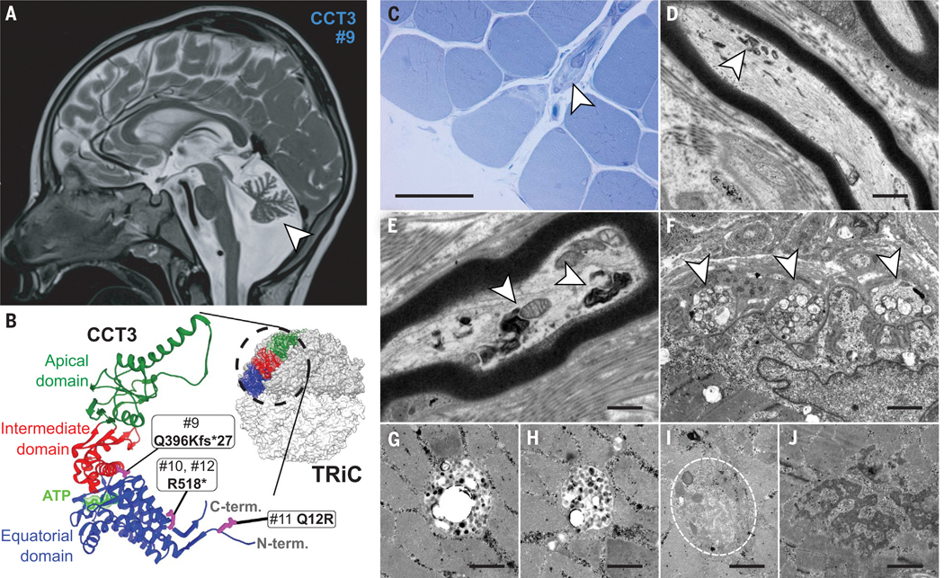
Malformations of the brain are common and vary in severity, from negligible to potentially fatal. Their causes have not been fully elucidated. Here, we report pathogenic variants in the core protein-folding machinery TRiC/CCT in individuals with brain malformations, intellectual disability, and seizures. The chaperonin TRiC is an obligate hetero-oligomer, and we identify variants in seven of its eight subunits, all of which impair function or assembly through different mechanisms. Transcriptome and proteome analyses of patient-derived fibroblasts demonstrate the various consequences of TRiC impairment. The results reveal an unexpected and potentially widespread role for protein folding in the development of the central nervous system and define a disease spectrum of "TRiCopathies."


Conflict of interest statement

Competing interests: The authors declare no competing interests.

Figures

Fig. 1.
Fig. 1.. CCT3 variants in human developmental disorder.
(A) MRI of individual #9 at age 4 years and 5 months showing hypoplastic cerebellar vermis (arrowhead) and hypomyelination of the white matter. (B) The heterozygous variants of the individuals (#9–#12) in CCT3 are indicated and locate across the different operational domains of the subunit. The apical domain of CCT3 (green) at the tip of the ring has a substrate recognition site and the lid-forming loop, the intermediate domain (red) controls ATP hydrolysis and thus the movement of the apical domain, and the equatorial domain (blue) includes the ATP-binding site (light green). Protein substrates of the complex are encapsulated in a folding chamber formed by TRiC to assist ATP-dependent folding of proteins. CCT3 is highlighted in the upper octameric ring of TRiC. The entire 16-mer complex is formed by two octameric rings. (C to J) Muscle biopsy findings. (C) Light microscopy of toluidine-blue–stained semithin section showing the spectrum of muscle fiber calibers. Arrowhead: intramuscular nerve fascicle containing myelinated nerve fibers. Scale bar, 40 μm. (D) Intramuscular nerve fiber showing intraaxonal accumulation of abnormal autophagic material (arrowhead). Electron micrograph (EM); scale bar, 2 μm. (E) In the intramuscular nerve fiber, the abnormal intraaxonal autophagic material (arrowheads) is associated with mitochondria. EM; scale bar, 1 μm. (F) Motor end plate displaying a paucity of synaptic folds and swelling of mitochondria in axon endings (arrowheads). EM; scale bar, 1 μm. (G and H) Two membrane-bound intermyofibrillar vacuoles containing globular and granular material of variable osmophilia. EM; scale bars, 600 nm. (I) intermyofibrillar accumulation of degenerating mitochondria and of vesicular material (dashed circle). EM; scale bar, 500 nm. (J) Representation of Z-band material. EM; scale bar, 1 μm.
Fig. 2.
Fig. 2.. Functional consequences of CCT3 alterations in different species.
(A) Conservation of the missense variant (individual #11) and position of the C-terminal loss-of-function variants (individual #9, #10, and #12) in different species. Single-letter abbreviations for the amino acid residues are as follows: A, Ala; C, Cys; D, Asp; E, Glu; F, Phe; G, Gly; H, His; I, Ile; K, Lys; L, Leu; M, Met; N, Asn; P, Pro; Q, Gln; R, Arg; S, Ser; T, Thr; V, Val; W, Trp; and Y, Tyr. (B) Functional studies of the cct3 variants by yeast plasmid-shuffling assays. (C) Representative brightfield images of C. elegans phenotype 48 hours after egg laying. Scale bars, 100 μm. (D) Actin staining in the intestine of cct-3 −/− and cct-3 Q14R/Q14R animals. Maximum intensity images of mCherry::ACT-5 in the intestine of WT (cct-3 +/+), heterozygous (cct-3 +/−), and homozygous (cct-3 −/−) L2 stage animals. Scale bars, 50 μm. (E) Quantification of cytoplasmic mCherry::ACT-5 puncta in adult worms. (F) Lethality in the presence of a green fluorescent protein (GFP)–β-tubulin fusion protein after heat shock. Shown are relative survival rates after heat shock at 37°C for 2 hours. Five independent biological replicates were combined for each genotype. Differences between groups were determined using ordinary one-way analysis of variance (ANOVA) followed by a Tukey multiple-comparisons test. ns, not significant; *P < 0.05; **P < 0.01; ****P < 0.0001 (G) Danio rerio cct3 knockout animals. Overview images of cct3 WT (cct3+/+) and homozygous knockout (cct3−/−) zebrafish at 4 days post fertilization (4 dpf) are shown. Scale bar, 1 mm. (H) Cerebellum of cct3+/+ and cct3−/− animals at 4 days dpf. Whole-mount immunohistochemistry of vglut1 as a marker of cerebellar granule cells (magenta) and pvalb7 as a marker of Purkinje cells (green). Scale bars, 40 μm. (I) F-actin was labeled using TRITC-conjugated phalloidin. Images of the hindbrains of WT zebrafish at 3 dpf (n = 5) and cct3 mutants (n = 4). Representative single planes are shown. Scale bar, 10 μm. The examined hindbrain area is marked with a red box in the diagram of the zebrafish larva (right).
Fig. 3.
Fig. 3.. Brain phenotype and cellular consequences of TRiC dysfunction.
(A to J) MRI images from affected individuals with CCT gene variants. (A) Individual age 29 years with heterotopia at the posterior horn of right lateral ventricle (arrowheads). (B) Child age 12 years with bilateral perisylvian polymicrogyria with frontal and parietal extension. (C) Child age 9 years with asymmetrical bilateral polymicrogyria, most severe over the right frontoparietal hemisphere (arrowheads in left and right panels). Mild findings perisylvian in the left hemisphere (arrowheads, middle). (D) Child age 14 years with bilateral symmetrical polymicrogyria, frontoparietal and temporal. (E) Child age 4 years with cerebellar atrophy and hypoplastic cerebellar vermis (arrowheads, right), hypomyelination of white matter, especially periventricular and temporal (middle). (F) Child age 2 years with reduced supratentorial white matter volume with abnormal signal, and thinning of the corpus callosum (middle). Reduced volume of the thalami bilaterally, with mild associated abnormal signal. Mildly small optic chiasm and optic nerves (left). (G) Individual with bilateral temporoparietal polymicrogyria (middle) and suspected dysplasia of posterior insular cortex (left) as well as hypogenesis of the corpus callosum (right), and hypoplasia of both cerebellar hemispheres and pons (middle). (H) Child age 2 years with cerebellar atrophy (arrowheads). (I) Child age 3 years with bilateral polymicrogyria. Extensive findings over the left hemisphere frontoparietal and temporal (arrowheads in left and right panels) and over the posterior end of sylvian fissure in the right hemisphere (arrow, middle panel). (J) Individual with extensive right hemispheric polymicrogyria, frontoparietal and temporal. (K) 16-mer TRiC complex with position of the CCT variants indicated in pink. (L) ATP-dependent protein folding via two octameric double rings, which build the folding chamber. (M) Pathogenic variants of all subunits projected onto a single prototype CCT primary structure (“panCCT”) with the three protein domains highlighted. (N) Structural modeling of the NSL variants. (O) Functional studies of missense mutations in CCT subunits in yeast. (P) High conservation of the NSL. (Q) Maximum intensity projection of actin aggregates in C. elegans harboring the orthologous CCT1-P38L human variant (cct-1 P42L) and CCT7-E379K human variant (cct-7 E377K), respectively. Scale bars, 50 μm (R) Quantification of actin aggregates ****P < 0.0001.
Fig. 4.
Fig. 4.. Downstream effects of human TRiC dysfunction.
(A to C) Transcriptome and proteome analysis in patient-derived fibroblast cell lines. Differences between groups were determined using t test and a P value ≤ 0.05. Volcano plots show the differentially expressed genes (B) and proteins (C) in the patient-derived cell lines (CCT3 #9, CCT5 #14, CCT6 #15, CCT8 #21) compared to healthy controls. Dashed lines indicate the thresholds for log2 fold change (≥0.585) and significance level (P ≤ 0.05). Up-regulated genes are shown in yellow, whereas down-regulated genes are colored in blue. Genes with expression changes common to all four genotypes (≥1.5-fold and with a P value ≤ 0.05) are labeled by gene or protein name. (D and E) Expression changes of all TRiC subunits as well as the genes and proteins of the actin and tubulin family are shown per genotype. (F and G) Pathway analysis of combined differentially regulated proteins from the patient-derived cells reveals up-regulated (F) and down-regulated (G) pathways.

Comment in

References

    1. Li Y. et al. , JAMA Neurol. 78, 578–587 (2021). - PMC - PubMed
    1. Klingler E, Francis F, Jabaudon D, Cappello S, Science 371, eaba4517 (2021). - PubMed
    1. Severino M. et al. , Brain 143, 2874–2894 (2020). - PMC - PubMed
    1. Oegema R. et al. , Nat. Rev. Neurol 16, 618–635 (2020). - PMC - PubMed
    1. Akula SK et al. , JAMA Neurol. 80, 980–988 (2023). - PMC - PubMed

Publication types

MeSH terms

LinkOut - more resources