. 2025 Apr;12(2):1386-1397.
doi: 10.1002/ehf2.15152. Epub 2024 Oct 31.

Semaglutide normalizes increased cardiomyocyte calcium transients in a rat model of high fat diet-induced obesity

Affiliations

Semaglutide normalizes increased cardiomyocyte calcium transients in a rat model of high fat diet-induced obesity

Vasco Sequeira et al. ESC Heart Fail. 2025 Apr.

Abstract

Aims: Obesity increases the risk of heart failure with preserved (HFpEF), but not reduced ejection fraction (HFrEF). The glucagon-like peptide-1 receptor agonist (GLP-1-RA) semaglutide improves outcome of patients with obesity with or without HFpEF, while GLP-1-RAs were associated with adverse outcome in patients with HFrEF. Here, we investigate the effect of in vivo treatment with semaglutide on excitation-contraction coupling in a rat model of obesity.

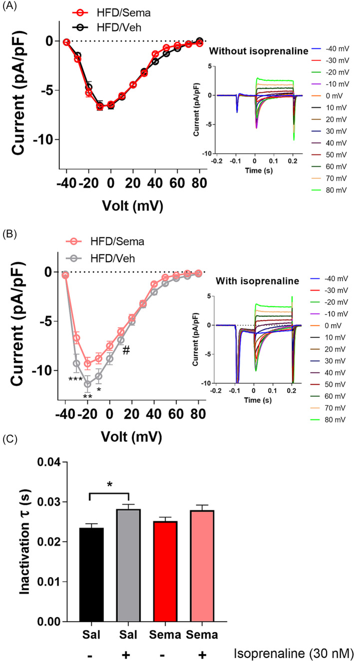
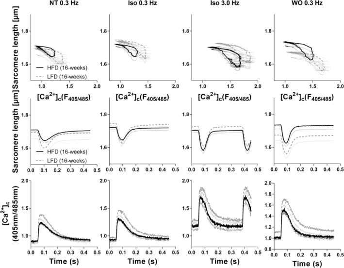
Methods and results: Rats received high-fat/high-fructose diet for 8 weeks and were then randomized to semaglutide (HFD/Sema) or vehicle (HFD/Veh) for another 8 weeks, during which they could choose between HFD and a low-fat/high-fructose diet (LFD). Control rats received either standard chow (CON), HFD or LFD only, without treatment. After 16 weeks, sarcomere shortening and cytosolic Ca2+ concentrations ([Ca2+]c) were determined in isolated cardiomyocytes. Compared with CON, HFD/Veh increased the amplitude of [Ca2+]c transients and systolic sarcomere shortening in absence or presence of β-adrenergic stimulation, which was reversed by HFD/Sema. Caffeine-induced sarcoplasmic reticulum (SR) Ca2+ release and L-type Ca2+ channel (LTCC) currents were reduced by HFD/Sema versus HFD/Veh, while SR Ca2+ ATPase activity remained unaffected. Compared with HFD, LFD increased [Ca2+]c transients and sarcomere shortening further despite similar effects on body weight.

Conclusions: While HFD increased cardiomyocyte [Ca2+]c transients and systolic sarcomere shortening, semaglutide normalized these alterations, mediated by reduced SR Ca2+ load and LTCC currents. Because increased LTCC currents were previously traced to cardiac hypertrophy, these effects may explain why GLP-1-RAs provide benefits for patients with obesity with or without HFpEF, but rather adverse outcome in HFrEF.

Keywords: Excitation‐contraction coupling; Glucagon‐like peptide agonists; Heart failure; Obesity; Semaglutide.


Conflict of interest statement

C.M. received speaker or advisory honoraria from Bristol Myers Squibb, Boehringer Ingelheim, AstraZeneca, Servier, Amgen, NovoNordisk, Bayer, Novartis, Edwards and Berlin Chemie. U.D. has received speaker honoraria of Alnylam, Recordati Rare Diseases and MSD. All other authors have declared that no conflict of interest exists.

Figures

Figure 1
Figure 1
Overview of the experimental design and the different treatment groups. Rats in the HFD/Sema group (n = 8) were fed a high‐fat, high‐fructose diet (HFD) for 8 weeks, followed by 8 weeks of treatment with subcutaneous semaglutide (120 μg/kg/day); during this treatment period, they had access to either the HFD or a low‐fat diet (LFD). The HFD/Veh group (n = 8) was managed similarly to the HFD/Sema group, except they received saline instead of semaglutide. Rats in the HFD group (n = 3) were given the high‐fat, high‐fructose diet, and the LFD group (n = 3) received the low‐fat diet for the entire duration of 16 weeks. Control rats (CON, n = 5) were maintained on a standard chow diet throughout the 16 weeks. Created with BioRender.com.
Figure 2
Figure 2
Sarcomeric and cytosolic Ca2+ dynamics transients in cardiomyocytes from controls (CON), non‐treated (HFD/Veh), and semaglutide‐treated high‐fat, high‐fructose diet (HFD) rat hearts. Cardiomyocytes were paced at 0.3 Hz under normal Tyrode conditions (NT), with 30 nM isoprenaline (Iso) at 0.3 Hz and 3 Hz, and during the washout (WO) phase at 0.3 Hz. (Top panels) Sarcomere length plotted to intracellular [Ca2+]c. (Mid panels) Diastolic and systolic sarcomere lengths. (Bottom panels) Diastolic and systolic intracellular [Ca2+]c. The washout (WO) phase refers to the time following the cessation of 3 Hz stimulation, still in the presence of isoprenaline, with stimulation resumed at 0.3 Hz frequency for 240 seconds.
Figure 3
Figure 3
Sarcomeric and cytosolic Ca2+ dynamics in cardiomyocytes from controls (CON), and non‐treated (HFD/Veh) and semaglutide‐treated high‐fat, high‐fructose diet (HFD) rat hearts. Cardiomyocytes were paced at 0.3 Hz and exposed to physiological stress conditions with 30 nM Isoprenaline and an increased stimulation rate to 3 Hz for 3 min. Panel (A) displays diastolic and systolic sarcomere lengths. Panel (B) illustrates percentage of sarcomere shortening. Panel (C) shows time to 50% and 90% sarcomere re‐lengthening. Panel (D) presents diastolic and systolic intracellular [Ca2+]c. Panel (E) illustrates the [Ca2+]c transient amplitude. Panel (F) shows the time to (TT) 50% and 90% [Ca2+]c decay. Control rat cardiomyocytes (n = 51 cardiomyocytes from n = 3 hearts). HFD/Sema cardiomyocytes were isolated from HFD rats treated with semaglutide (n = 120 cardiomyocytes from n = 5 hearts). HFD/Veh cardiomyocytes were isolated from HFD rats administered saline (n = 100 cardiomyocytes from n = 3 hearts). Washout phase (WO) refers to the time following the cessation of 3 Hz stimulation, with stimulation resumed at 0.3 Hz frequency. Statistical significance: # P < 0.05 HFD/Sema versus HFD/Veh (two‐way ANOVA); *P < 0.05 CON versus HFD/Veh (two‐way ANOVA); τ < 0.05 CON versus HFD/Sema (two‐way ANOVA).
Figure 4
Figure 4
Sarcoplasmic reticulum (SR) Ca2+ load in cardiomyocytes from non‐treated (HFD/Veh) and semaglutide‐treated high‐fat, high‐fructose diet (HFD) hearts. (A) Original tracings showcase caffeine‐induced increases in cytosolic [Ca2+]c, serving as an indicator of SR Ca2+ load, at 1 Hz pacing and 10 mM caffeine. Panel (B) Displays the amplitude of [Ca2+]c changes (Δ[Ca2+]c). Panel (C) Illustrates the tau (τ) of [Ca2+]c removal at 1 Hz, indicative of SR Ca2+‐ATPase (SERCA) activity, and during caffeine application, representing Na+/Ca2+‐exchanger (NCX) activity. Panel (D) reveals the time to peak (TPP) [Ca2+]c at 1 Hz and during caffeine application. HFD/Sema cardiomyocytes were isolated from HFD rats treated with semaglutide (n = 42 cardiomyocytes from n = 3 hearts). HFD/Veh cardiomyocytes were isolated from HFD rats administered saline (n = 49 cardiomyocytes from n = 3 hearts). Sal, Saline; Sema, Semaglutide. Statistical significance: #P < 0.05 (one‐way ANOVA); *P < 0.05, **P < 0.01, ****P < 0.0001.
Figure 5
Figure 5
L‐Type Ca2+ channel activity through patch‐clamp electrophysiology. Panel (A) presents baseline activity of L‐Type Ca2+ channel, and (B) its activity during the application of 30 nM Isoprenaline, with representative protocols illustrated in the insets. Panel (C) outlines the Ca2+‐induced inactivation tau (τ) kinetics of the L‐type Ca2+ channel. HFD/Sema cardiomyocytes were isolated from HFD rats treated with semaglutide (n = 33 cardiomyocytes from n = 3 hearts). HFD/Veh cardiomyocytes were isolated from HFD rats administered saline (n = 29 cardiomyocytes from n = 3 hearts). Sal, Saline; Sema, Semaglutide. Statistical significance: #P < 0.05 (two‐way ANOVA); *P < 0.05, **P < 0.01, ***P < 0.001, ****P < 0.0001.
Figure 6
Figure 6
Sarcomeric and cytosolic Ca2+ dynamics in cardiomyocytes from low‐fat (LFD) and high‐fat, high‐fructose diet (HFD) rat hearts. Cardiomyocytes were paced at 0.3 Hz and exposed to physiological stress conditions with 30 nM Isoprenaline and an increased stimulation rate to 3 Hz for 3 min. Panel (A) displays diastolic and systolic sarcomere lengths. Panel (B) illustrates percentage of sarcomere shortening. Panel (C) shows time to 50% and 90% sarcomere re‐lengthening. Panel (D) presents diastolic and systolic intracellular [Ca2+]c. Panel (E) illustrates the [Ca2+]c transient amplitude. Panel (F) shows the time to (TT) 50% and 90% [Ca2+]c decay. LFD (16 weeks), cardiomyocytes isolated from rats maintained on a low‐fat diet for 16 weeks (n = 79 cardiomyocytes from n = 3 hearts). HFD (16 weeks), cardiomyocytes isolated from rats subjected to a high‐fat, high‐fructose diet for 16 weeks (n = 70 cardiomyocytes from n = 3 hearts). WO, washout phase, refers to the time following the cessation of 3 Hz stimulation, with stimulation resumed at 0.3 Hz frequency. Statistical significance: # P < 0.05 HFD versus LFD (two‐way ANOVA).
Figure 7
Figure 7
Sarcomeric and cytosolic Ca2+ dynamics transients in cardiomyocytes from low‐fat (LFD) and high‐fat, high‐fructose diet (HFD) rat hearts. Cardiomyocytes were paced at 0.3 Hz under normal Tyrode conditions (NT), with 30 nM isoprenaline (Iso) at 0.3 Hz and 3 Hz, and during the washout (WO) phase at 0.3 Hz. (Top panels) Sarcomere length plotted to intracellular [Ca2+]c. (Mid panels) Diastolic and systolic sarcomere lengths. (Bottom panels) Diastolic and systolic intracellular [Ca2+]c. The washout (WO) phase refers to the time following the cessation of 3 Hz stimulation, still in the presence of isoprenaline, with stimulation resumed at 0.3 Hz frequency for 240 s.

References

    1. Collaborators GBDO , Afshin A, Forouzanfar MH, Reitsma MB, Sur P, Estep K, et al. Health effects of overweight and obesity in 195 countries over 25 years. N Engl J Med 2017;377:13‐27. doi:10.1056/NEJMoa1614362 - DOI - PMC - PubMed
    1. Pandey A, LaMonte M, Klein L, Ayers C, Psaty BM, Eaton CB, et al. Relationship between physical activity, body mass index, and risk of heart failure. J Am Coll Cardiol 2017;69:1129‐1142. doi:10.1016/j.jacc.2016.11.081 - DOI - PMC - PubMed
    1. Adamson C, Kondo T, Jhund PS, de Boer RA, Cabrera Honorio JW, Claggett B, et al. Dapagliflozin for heart failure according to body mass index: the DELIVER trial. Eur Heart J 2022;43:4406‐4417. doi:10.1093/eurheartj/ehac481 - DOI - PMC - PubMed
    1. Borlaug BA, Kane GC, Melenovsky V, Olson TP. Abnormal right ventricular‐pulmonary artery coupling with exercise in heart failure with preserved ejection fraction. Eur Heart J 2016;37:3293‐3302. doi:10.1093/eurheartj/ehw241 - DOI - PMC - PubMed
    1. Rosch S, Kresoja K‐P, Besler C, Fengler K, Schöber AR, Mv R, et al. Characteristics of heart failure with preserved ejection fraction across the range of left ventricular ejection fraction. Circulation 2022;146:506‐518. doi:10.1161/CIRCULATIONAHA.122.059280 - DOI - PubMed