. 2024 Nov;56(11):2465-2474.
doi: 10.1038/s12276-024-01344-6. Epub 2024 Nov 1.

The MCP-3/Ccr3 axis contributes to increased bone mass by affecting osteoblast and osteoclast differentiation

Affiliations

The MCP-3/Ccr3 axis contributes to increased bone mass by affecting osteoblast and osteoclast differentiation

Jung Ha Kim et al. Exp Mol Med. 2024 Nov.

Abstract

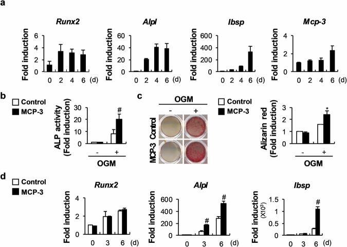
Several CC subfamily chemokines have been reported to regulate bone metabolism by affecting osteoblast or osteoclast differentiation. However, the role of monocyte chemotactic protein 3 (MCP-3), a CC chemokine, in bone remodeling is not well understood. Here, we show that MCP-3 regulates bone remodeling by promoting osteoblast differentiation and inhibiting osteoclast differentiation. In a Ccr3-dependent manner, MCP-3 promoted osteoblast differentiation by stimulating p38 phosphorylation and suppressed osteoclast differentiation by upregulating interferon beta. MCP-3 increased bone morphogenetic protein 2-induced ectopic bone formation, and mice with MCP-3-overexpressing osteoblast precursor cells presented increased bone mass. Moreover, MCP-3 exhibited therapeutic effects by abrogating receptor activator of nuclear factor kappa-B ligand-induced bone loss. Therefore, MCP-3 has therapeutic potential for diseases involving bone loss due to its positive role in osteoblast differentiation and negative role in osteoclast differentiation.


Conflict of interest statement

Competing interests: The authors declare no competing interests.

Figures

Fig. 1
Fig. 1. MCP-3 promotes osteoblast differentiation and function.
a Osteoblasts were cultured in osteogenic medium (OGM). The relative mRNA levels of the indicated genes were determined via RT‒qPCR (n = 3). bd Osteoblasts were cultured in OGM with or without MCP-3. b After culture for three days, the cells were lysed and subjected to alkaline phosphatase (ALP) activity measurement (n = 3). c After culture for six days, the cells were stained with Alizarin Red and quantified via extraction (n = 3). d The relative mRNA levels of the indicated genes were determined via RT‒qPCR (n = 3). The data are presented as the means ± SDs of triplicate samples. # and * indicate p < 0.05 and < 0.01, respectively, vs. the control.
Fig. 2
Fig. 2. In osteoblasts, MCP-3 regulates nodule formation by increasing p38 phosphorylation in a Ccr3-dependent manner.
a Serum-starved osteoblasts pretreated with or without MCP-3 for 60 min were stimulated with bone morphogenetic protein 2 (BMP2) for the indicated times, followed by western blot analysis of the indicated proteins (n = 3). b, c Osteoblasts were cultured in OGM with or without MCP-3 and SB203580. b Cultured cells were stained with Alizarin Red and quantified via extraction (n = 3). c The relative mRNA levels of the indicated genes were determined via RT‒qPCR (n = 3). d Osteoblasts were cultured in OGM, and the relative mRNA levels of the indicated genes were determined via RT‒qPCR (n = 3). e Osteoblasts were cultured in the presence or absence of MCP-3, and the relative Ccr3 mRNA level was determined via RT‒qPCR (n = 3). f Osteoblasts were transfected with Con-siRNA or Ccr3-siRNA and then cultured in OGM. The relative mRNA levels of the indicated genes were determined via RT‒qPCR (n = 3). g, h Osteoblasts were transfected with Con-siRNA or Ccr3-siRNA and cultured with OGM in the presence or absence of MCP-3. g Relative Ccr3 mRNA levels were determined via RT‒qPCR (n = 3). h Cultured cells were stained with Alizarin Red and quantified via extraction (n = 3). i After transfection with Con-siRNA or Ccr3-siRNA, osteoblasts were serum-starved, pretreated with MCP-3, stimulated with BMP2, and then subjected to western blot analysis of the indicated proteins (n = 3). The data are presented as the means ± SDs of triplicate samples. #, *, and ** indicate p < 0.05, < 0.01, and < 0.001, respectively, vs. the control.
Fig. 3
Fig. 3. MCP-3 inhibits osteoclast differentiation.
a Bone marrow-derived macrophages (BMMs) were differentiated with macrophage colony-stimulating factor (M-CSF) and receptor activator of NF-κB ligand (RANKL). The relative mRNA levels of the indicated genes were determined via RT‒qPCR (n = 3). b BMMs were differentiated with M-CSF and RANKL and treated with the indicated MCP-3 concentrations during osteoclast differentiation. The cultured cells were stained with tartrate-resistant acid phosphatase (TRAP), and TRAP-positive cells were counted (n = 3; scale bar: 200 µm). c BMMs were cultured with M-CSF and treated with the indicated MCP-3 concentrations, followed by an MTT assay after three days. d, e BMMs were differentiated with M-CSF and RANKL with or without MCP-3. d The relative mRNA levels of the indicated genes were determined via RT‒qPCR (n = 3). e The indicated proteins were assayed via western blot analysis (n = 3). The data are presented as the means ± SDs of triplicate samples. #, *, and ** indicate p < 0.05, < 0.01, and < 0.001, respectively, vs. the control.
Fig. 4
Fig. 4. MCP-3 inhibits osteoclast differentiation via Ccr3-dependent interferon beta (IFNβ) upregulation.
a BMMs were differentiated with M-CSF and RANKL, and the relative mRNA levels of the indicated genes were determined via RT‒qPCR (n = 3). b BMMs were cultured with M-CSF with or without MCP-3. The relative Ccr3 mRNA level was determined via RT‒qPCR (n = 3). c Con-siRNA- or Ccr3-siRNA-transfected BMMs were cultured with M-CSF, and the relative Ccr3 mRNA level was determined via RT‒qPCR (n = 3). d, e BMMs were transfected with Con-siRNA or Ccr3-siRNA and then cultured with M-CSF and RANKL. d Cultured cells were stained with TRAP, and TRAP-positive cells were counted (n = 3; scale bar: 200 µm). e The relative mRNA levels of the indicated genes were determined via RT‒qPCR (n = 3). f BMMs were transfected with Con-siRNA or Ccr3-siRNA and cultured with M-CSF and RANKL, with or without MCP-3. The cultured cells were stained with TRAP, and TRAP-positive cells were counted (n = 3; scale bar: 200 µm). g BMMs were cultured with M-CSF with or without MCP-3, and the relative Ifnb mRNA level was determined via RT‒qPCR (n = 3). h BMMs were cultured with M-CSF and RANKL, with or without MCP-3 and an IFNβ-blocking antibody. The cultured cells were stained with TRAP, and TRAP-positive cells were counted (n = 3; scale bar: 200 µm). i BMMs were transfected with Con-siRNA or Ccr3-siRNA and then cultured with M-CSF and RANKL. The relative mRNA levels of the indicated genes were determined via RT‒qPCR (n = 3). The data are presented as the means ± SDs of triplicate samples. #, *, and ** indicate p < 0.05, < 0.01, and < 0.001, respectively, vs. the control.
Fig. 5
Fig. 5. Transgenic mice overexpressing osteoblast-specific MCP-3 presented increased long-bone mass.
ac Bone marrow stromal cells from MCP-3 transgenic mice or their wild-type littermates were cultured with or without OGM. a ALP activity was measured (n = 3). b Cultured cells were stained with Alizarin Red and quantified via extraction (n = 3). c The relative mRNA levels of the indicated genes were determined via RT‒qPCR (n = 3). d, e Microcomputed tomography (µCT) and histological analyses of long bones from MCP-3 transgenic mice or their wild-type littermates. d Representative µCT 3D images of femurs from MCP-3 transgenic mice or their wild-type littermates. The bone volume/tissue volume (BV/TV), trabecular thickness (Tb.Th), trabecular separation (Tb.Sp), trabecular number (Tb.N), cortical bone volume/tissue volume (Corti BV/TV), and cortical thickness (Corti Th) were determined via µCT (n = 7 or 10). e Hematoxylin and eosin- and TRAP-stained images of tibiae from MCP-3 transgenic mice or their wild-type littermates. Osteoblasts and osteoclasts were quantified via histological analyses (n = 4 or 5). The data are presented as the means ± SDs. #, *, and ** indicate p < 0.05, < 0.01, and < 0.001, respectively, vs. the control.
Fig. 6
Fig. 6. MCP-3 increases bone formation in vivo.
a Collagen sponges soaked with BMP2, with or without MCP-3, were subcutaneously implanted on top of the dorsal back. Ectopic bones were biopsied and subjected to µCT analyses. Representative µCT 3D ectopic bone images are shown. The bone volume and bone area were determined via µCT (n = 5). b Ten and two days before euthanasia, MCP-3 transgenic mice and their wild-type littermates were intraperitoneally injected with calcein green. Representative calcein double-labeled images are shown. The cortical bone mineralizing surface/bone surface (Ct. MS/BS), cortical bone mineral apposition rate (Ct. MAR), cortical bone formation rate (Ct. BFR), trabecular mineralizing surface/bone surface (Tb. MS/BS), trabecular mineral apposition rate (Tb. MAR), and trabecular bone formation rate (Tb. BFR) were assessed (n = 4 or 6, scale bar: 50 µm). c Mice were intraperitoneally injected with PBS or MCP-3 one day before RANKL injection. RANKL and PBS or RANKL and MCP-3 were injected daily (simultaneously) for three days. Representative 3D femur images are shown. The bone volume/tissue volume (BV/TV), trabecular thickness (Tb.Th), trabecular separation (Tb.Sp), and trabecular number (Tb.N) were assessed via µCT (n = 4). The data are presented as the means ± SDs. #, *, and ** indicate p < 0.05, < 0.01, and < 0.001, respectively, vs. the control.
Fig. 7
Fig. 7. MCP-3 is involved in irisin-regulated osteoblast and osteoclast differentiation.
a Osteoblasts were cultured in OGM with or without irisin. The relative mRNA levels of the indicated genes were determined via RT‒qPCR (n = 3). b Osteoblasts were transfected with Con-siRNA or Mcp-3-siRNA. The relative Mcp-3 mRNA level was determined via RT‒qPCR (n = 3). c Osteoblasts were transfected with Con-siRNA or Mcp-3-siRNA and cultured with OGM, with or without irisin. Cultured cells were stained with Alizarin Red and quantified via extraction (n = 3). d BMMs were differentiated with M-CSF and RANKL, with or without irisin. The relative mRNA levels of the indicated genes were determined via RT‒qPCR (n = 3). e BMMs were transfected with Con-siRNA or Mcp-3-siRNA and then cultured with M-CSF. The relative Mcp-3 mRNA level was determined via RT‒qPCR (n = 3). f BMMs were transfected with Con-siRNA or Mcp-3-siRNA and cultured with M-CSF and RANKL, with or without irisin. Cultured cells were stained with TRAP, and TRAP-positive cells were counted (n = 3; scale bar: 200 µm). The data are presented as the means ± SDs of triplicate samples. #, *, and ** indicate p < 0.05, < 0.01, and < 0.001, respectively, vs. the control.

References

    1. Kim, J. H. & Kim, N. Bone cell communication factors provide a new therapeutic strategy for osteoporosis. Chonnam Med J.56, 94–98 (2020). - PMC - PubMed
    1. Lee, H. Y., Jung, J. E. & Yim, M. Iris Koreana NAKAI inhibits osteoclast formation via p38-mediated nuclear factor of activated T cells 1 signaling pathway. J. Bone Metab.30, 253–262 (2023). - PMC - PubMed
    1. Kim, K., Han, J. E., Lee, K. B. & Kim, N. LIM homeobox transcription factor 1-β expression is upregulated in patients with osteolysis after total ankle arthroplasty and inhibits receptor activator of nuclear factor-κB ligand-induced osteoclast differentiation in vitro. J. Bone Metab.29, 165–174 (2022). - PMC - PubMed
    1. Wintges, K. et al. Impaired bone formation and increased osteoclastogenesis in mice lacking chemokine (C-C motif) ligand 5 (Ccl5). J. Bone Min. Res28, 2070–2080 (2013). - PubMed
    1. Sabatini, M., Boyce, B., Aufdemorte, T., Bonewald, L. & Mundy, G. R. Infusions of recombinant human interleukins 1 alpha and 1 beta cause hypercalcemia in normal mice. Proc. Natl. Acad. Sci. USA85, 5235–5239 (1988). - PMC - PubMed

LinkOut - more resources