Observational Study
. 2024 Oct 31;16(1):242.
doi: 10.1186/s13195-024-01603-8.

Perivascular space enlargement accelerates in ageing and Alzheimer's disease pathology: evidence from a three-year longitudinal multicentre study

Inga Menze #  1   2 Jose Bernal #  3   4   5   6 Pinar Kaya  3   7 Çağla Aki  3   7 Malte Pfister  7 Jonas Geisendörfer  7 Renat Yakupov  3   4 Roberto Duarte Coello  5   6 Maria D C Valdés-Hernández  5   6 Michael T Heneka  8 Frederic Brosseron  9 Matthias C Schmid  9   10 Wenzel Glanz  3   4 Enise I Incesoy  3   4   11 Michaela Butryn  3   4 Ayda Rostamzadeh  12 Dix Meiberth  9   12 Oliver Peters  13   14 Lukas Preis  14 Dominik Lammerding  14 Daria Gref  14 Josef Priller  6   13   15   16 Eike J Spruth  13   15 Slawek Altenstein  13   15 Andrea Lohse  15 Stefan Hetzer  17 Anja Schneider  9   18 Klaus Fliessbach  9   18 Okka Kimmich  9 Ina R Vogt  9 Jens Wiltfang  19   20   21 Claudia Bartels  20 Björn H Schott  19   20   22 Niels Hansen  20 Peter Dechent  23 Katharina Buerger  24   25 Daniel Janowitz  25 Robert Perneczky  24   26   27   28 Boris-Stephan Rauchmann  26   29   30 Stefan Teipel  31   32 Ingo Kilimann  31   32 Doreen Goerss  31   32 Christoph Laske  33   34 Matthias H Munk  33   35 Carolin Sanzenbacher  33 Petra Hinderer  33 Klaus Scheffler  36 Annika Spottke  9   37 Nina Roy-Kluth  9 Falk Lüsebrink  3 Katja Neumann  7 Joanna Wardlaw  5   6 Frank Jessen  9   12   38 Stefanie Schreiber #  3   7 Emrah Düzel #  3   4 Gabriel Ziegler #  3   4
Affiliations
Observational Study

Perivascular space enlargement accelerates in ageing and Alzheimer's disease pathology: evidence from a three-year longitudinal multicentre study

Inga Menze et al. Alzheimers Res Ther. .

Abstract

Background: Perivascular space (PVS) enlargement in ageing and Alzheimer's disease (AD) and the drivers of such a structural change in humans require longitudinal investigation. Elucidating the effects of demographic factors, hypertension, cerebrovascular dysfunction, and AD pathology on PVS dynamics could inform the role of PVS in brain health function as well as the complex pathophysiology of AD.

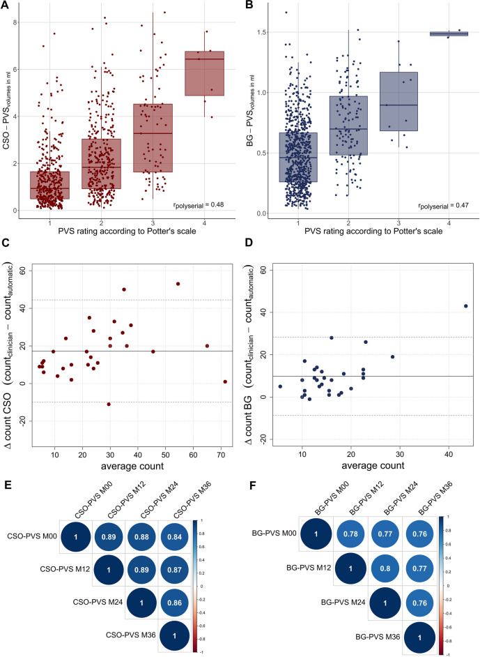
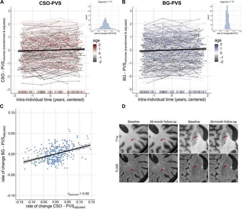
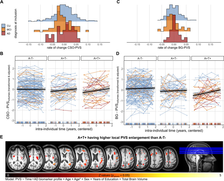
Methods: We studied PVS in centrum semiovale (CSO) and basal ganglia (BG) computationally over three to four annual visits in 503 participants (255 females; meanage = 70.78 ± 5.78) of the ongoing observational multicentre "DZNE Longitudinal Cognitive Impairment and Dementia Study" (DELCODE) cohort. We analysed data from subjects who were cognitively unimpaired (n = 401), had amnestic mild cognitive impairment (n = 71), or had AD (n = 31). We used linear mixed-effects modelling to test for changes of PVS volumes in relation to cross-sectional and longitudinal age, as well as sex, years of education, hypertension, white matter hyperintensities, AD diagnosis, and cerebrospinal-fluid-derived amyloid (A) and tau (T) status (available for 46.71%; A-T-/A + T-/A + T + n = 143/48/39).

Results: PVS volumes increased significantly over follow-ups (CSO: B = 0.03 [0.02, 0.05], p < 0.001; BG: B = 0.05 [0.03, 0.07], p < 0.001). PVS enlargement rates varied substantially across subjects and depended on the participant's age, white matter hyperintensities volumes, and amyloid and tau status. PVS volumes were higher across elderly participants, regardless of region of interest (CSO: B = 0.12 [0.02, 0.21], p = 0.017; BG: B = 0.19 [0.09, 0.28], p < 0.001). Faster BG-PVS enlargement related to lower baseline white matter hyperintensities volumes (ρspearman = -0.17, pFDR = 0.001) and was more pronounced in individuals who presented with combined amyloid and tau positivity versus negativity (A + T + > A-T-, pFDR = 0.004) or who were amyloid positive but tau negative (A + T + > A + T-, pFDR = 0.07). CSO-PVS volumes increased at a faster rate with amyloid positivity as compared to amyloid negativity (A + T-/A + T + > A-T-, pFDR = 0.021).

Conclusion: Our longitudinal evidence supports the relevance of PVS enlargement in presumably healthy ageing as well as in AD pathology. We further discuss the region-specific involvement of white matter hyperintensities and neurotoxic waste accumulation in PVS enlargement and the possibility of additional factors contributing to PVS progression. A comprehensive understanding of PVS dynamics could facilitate the understanding of pathological cascades and might inform targeted treatment strategies.

Trial registration: German Clinical Trials Register DRKS00007966. Registered 04.05.2015 - retrospectively registered, https://drks.de/search/en/trial/DRKS00007966 .

Keywords: Alzheimer’s disease; Alzheimer’s pathology; Enlarged perivascular spaces; Longitudinal analysis; Multicentre study; Virchow–Robin spaces.


Conflict of interest statement

The authors declare no competing interests.

Figures

Fig. 1
Fig. 1
Clinical validation and reliability using repeated measures. A-B Moderate polyserial correlations between PVS volumes and visual scores. C-D Bland–Altman plot comparing manual and computational PVS counts in the axial slice with the highest PVS burden in white matter and basal ganglia in a subset of 30 random subjects. Solid lines depict the mean difference and dotted lines depict the corresponding 95%-confidence intervals. E–F High Pearson’s correlations of CSO- and BG-PVS volumes across four annual time points suggest measurement stability. PVS volumes were Box-Cox transformed and z-scored to account for skewness and corrected for linear and quadratic age effects, years of education, sex and total intracranial volume
Fig. 2
Fig. 2
PVS enlargement over follow-ups and with advancing age in cognitively unimpaired subjects. A-B CSO- and BG-PVS volumes increased over time (CSO: B = 0.03 [95%-CI 0.02, 0.05], p < 0.001; BG: B = 0.05, 95%-CI [0.03, 0.07], p < 0.001). Histograms show respective distribution of rates of change in cognitively unimpaired individuals, using transformed data, corrected for effects of age, sex, education, total intracranial volume and regional volumes of WMH of presumed vascular origin. We Box-Cox transformed and z-scored PVS volumes. Plotted PVS volumes were furthermore adjusted for effects of sex, education and total intracranial volume. C Moderate, positive correlation of CSO- and BG-PVS change rates. Change rates were corrected for linear and quadratic age effects, sex, years of education, sbTIV as well as regional volumes of WMH of presumed vascular origin. D Within a three-year period, volumes of WMH of presumed vascular origin may emerge in the vicinity of PVS. Example images of a CU participant from two different axial slices
Fig. 3
Fig. 3
PVS enlargement is associated with AD biomarker profiles. PVS volumes in all plots were Box-Cox and z-transformed and adjusted for linear and quadratic age effects, sex, years of education, total intracranial volume and regional WMH of presumed vascular origin. A Histogram ridgeline plots of distribution of CSO-PVS rates of change across diagnostic groups. B Increase of CSO-PVS is dependent on the AD biomarker profile. Colour of individual trajectories corresponds to diagnosis at time of entry to DELCODE study. Subjects with A + T- or A + T + status show higher rates of change as compared to A-T-. C Histogram ridgeline plots of distribution of BG-PVS rates of change across diagnostic groups. D Increase of BG-PVS is dependent on the AD biomarker profile. Colour of individual trajectories corresponds to diagnosis at time of entry to DELCODE study. Subjects with A + T + status show higher rates of change as compared to A-T-. E Contrast image highlighting regions where PVS enlargement was more evident in A + T + vs. A-T- (puncorr < 0.05). We registered all PVS segmentation maps to a DELCODE-specific Multi-Brain (MB) toolbox template [58] and adjusted for local volume changes introduced by normalisation in PVS segmentation maps by modulation with Jacobian determinants [59, 60]. PVS maps were smoothed with Gaussian kernels (6 mm full width at half maximum). Model was aligned with regional marginal models [54] (PVS ~ Time*AT profile + Age + Age.2 + Sex + Years of Education + Total Brain Volume)

References

    1. Troili F, Cipollini V, Moci M, Morena E, Palotai M, Rinaldi V, et al. Perivascular unit: this must be the place. The anatomical crossroad between the immune, vascular and nervous system. Front Neuroanat. 2020;14. Available from: https://www.frontiersin.org/article/10.3389/fnana.2020.00017/full. - DOI - PMC - PubMed
    1. Wardlaw JM, Benveniste H, Nedergaard M, Zlokovic BV, Mestre H, Lee H, et al. Perivascular spaces in the brain: anatomy, physiology and pathology. Nat Rev Neurol. 2020;16(3):137–53. 10.1038/s41582-020-0312-z. - PubMed
    1. Del Brutto OH, Mera RM, Costa AF, Rumbea DA, Recalde BY, Del Brutto VJ. Patterns of progression of cerebral small vessel disease markers in older adults of Amerindian ancestry: a population-based, longitudinal prospective cohort study. Aging Clin Exp Res. 2022;34(11):2751–9. 10.1007/s40520-022-02223-8. - PMC - PubMed
    1. Francis F, Ballerini L, Wardlaw JM. Perivascular spaces and their associations with risk factors, clinical disorders and neuroimaging features: a systematic review and meta-analysis. Int J Stroke. 2019;14(4):359–71 Available from: 10.1177/1747493019830321. - DOI - PubMed
    1. Li Y, Kalpouzos G, Laukka EJ, Dekhtyar S, Bäckman L, Fratiglioni L, et al. Progression of neuroimaging markers of cerebral small vessel disease in older adults: A 6-year follow-up study. Neurobiol Aging. 2022;112:204–11 Available from: 10.1016/j.neurobiolaging.2022.01.006 . - PubMed

Publication types