. 2024 Oct 31;16(1):126.
doi: 10.1186/s13073-024-01400-w.

Circular RNA landscape in extracellular vesicles from human biofluids

Affiliations

Circular RNA landscape in extracellular vesicles from human biofluids

Jingjing Zhao et al. Genome Med. .

Abstract

Background: Circular RNAs (circRNAs) have emerged as a prominent class of covalently closed single-stranded RNA molecules that exhibit tissue-specific expression and potential as biomarkers in extracellular vesicles (EVs) derived from liquid biopsies. Still, their characteristics and applications in EVs remain to be unveiled.

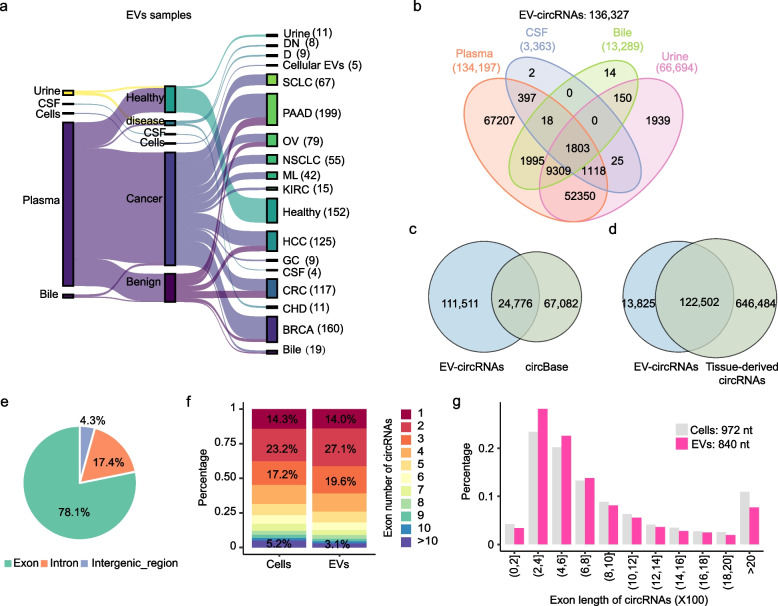
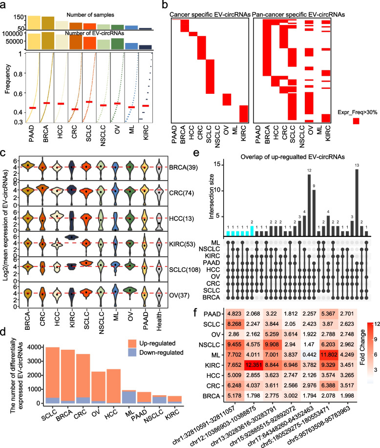
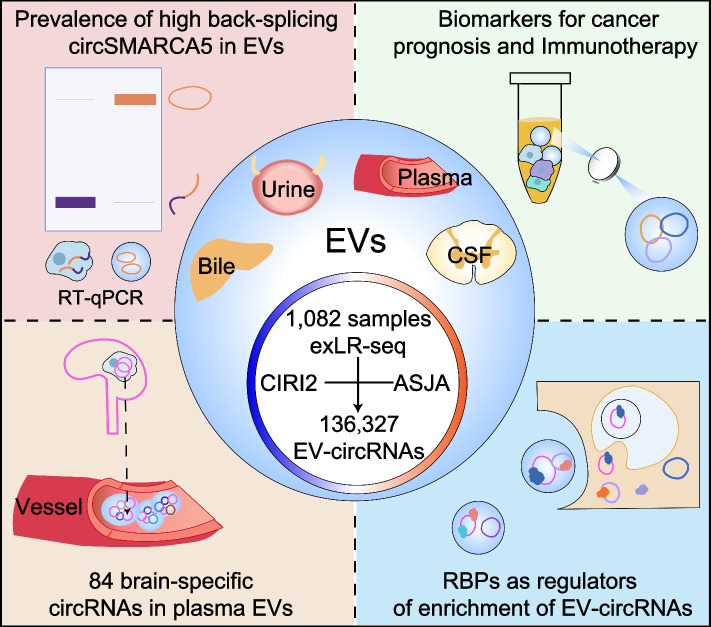
Methods: We performed a comprehensive analysis of EV-derived circRNAs (EV-circRNAs) using transcriptomics data obtained from 1082 human body fluids, including plasma, urine, cerebrospinal fluid (CSF), and bile. Our validation strategy utilized RT-qPCR and RNA immunoprecipitation assays, complemented by computational techniques for analyzing EV-circRNA features and RNA-binding protein interactions.

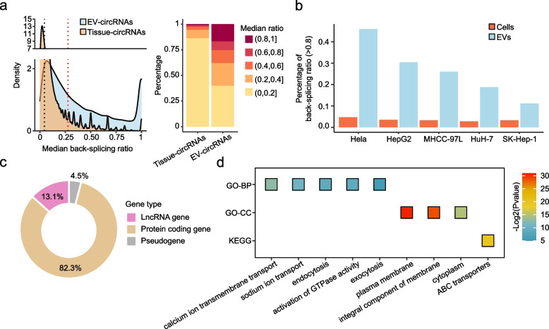
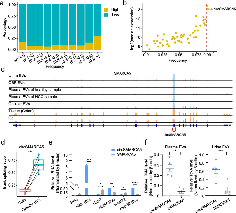
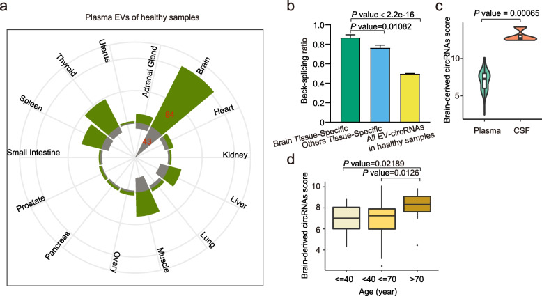
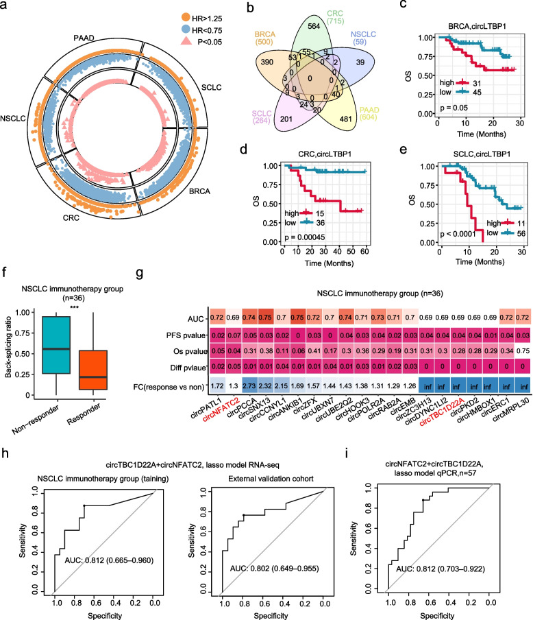
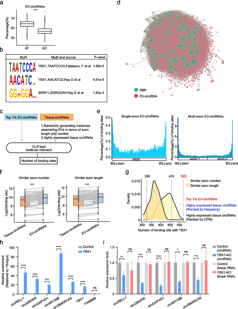
Results: We identified 136,327 EV-circRNAs from various human body fluids. Significantly, a considerable amount of circRNAs with a high back-splicing ratio are highly enriched in EVs compared to linear RNAs. Additionally, we discovered brain-specific circRNAs enriched in plasma EVs and cancer-associated EV-circRNAs linked to clinical outcomes. Moreover, we demonstrated that EV-circRNAs have the potential to serve as biomarkers for evaluating immunotherapy efficacy in non-small cell lung cancer (NSCLC). Importantly, we identified the involvement of RBPs, particularly YBX1, in the sorting mechanism of circRNAs into EVs.

Conclusions: This study unveils the extensive repertoire of EV-circRNAs across human biofluids, offering insights into their potential as disease biomarkers and their mechanistic roles within EVs. The identification of specific circRNAs and the elucidation of RBP-mediated sorting mechanisms open new avenues for the clinical application of EV-circRNAs in disease diagnostics and therapeutics.

Keywords: Biomarker; Cancer; Circular RNAs; Extracellular vesicles; RNA-binding proteins.


Conflict of interest statement

The authors declare no competing interests.

Figures

Fig. 1
Fig. 1
Characterization of circRNA atlas from 1082 EV samples by exLR-seq. a The figure represents the EV samples analyzed in our study, with the number in parentheses indicating the number of samples in each cohort. Extracellular vesicles, EVs; Breast cancer, BRCA; Colon cancer, CRC; Hepatocellular Carcinoma, HCC; Pancreatic Adenocarcinoma, PAAD; Ovarian cancer, OV; Coronary heart disease, CHD; Gastric cancer, GC; Kidney cancer, KIRC; Malignant lymphoma, ML; Non-small cell lung cancer, NSCLC; Small cell lung cancer, SCLC; Cerebrospinal fluid, CSF; Diabetes, D; Diabetic Nephropathy, DN. b Venn diagram showing the overlap of circRNAs detected in EVs from plasma, CSF, bile, and urine. c Overlap of circRNAs between EVs and circBase. d Overlap of circRNAs between EVs and tissue-derived circRNAs (circAltas 3.0). e Genomic origin of EV-circRNAs. f Distribution of circRNA exon numbers in cells (5 cell lines) and secreted EVs. g Exon length distribution of circRNAs in cells (5 cell lines) and secreted EVs.
Fig. 2
Fig. 2
High back-splicing ratio of circRNAs in EVs. a Density plot showing the distribution of the back-splicing ratio between EVs and tissues cohort, the bar plot displays the percentage of circRNAs in different back-splicing ratio intervals. The black dashed line represents the median back-splicing ratio in tissue, while the red line represents the median back-splicing ratio in EVs. b Bar plot showing the percentage of circRNAs with high back-splicing ratio in 5 cell lines and their secreted EVs. c Distribution of host genes for EV-circRNAs with a back-splicing ratio of 1 and median CPM >50. d Gene Ontology (GO) and Kyoto Encyclopedia of Genes and Genomes (KEGG) enrichment analysis for the host genes in Figure 2c
Fig. 3
Fig. 3
Identification of circSMARCA5 as EV marker. a Percentage of EV-circRNAs in different frequency ranges. High: back-splicing ratio>0.8. b The expression (median CPM) and frequency of EV-circRNAs with a high back-splicing ratio. c IGV plots showing the reads coverage of circSMARCA5. d Boxplot showing the back-splicing ratio of circSMARCA5 between cells and their secreted EVs. Each point represents a cell line. Paired T-test, two-sided, ***p < 0.001. e Relative expression of circSMARCA5 and SMARCA5 in cells and their secreted EVs. Statistical analysis was performed using unpaired Student’s t-tests. *p < 0.05; **p < 0.01; ***p < 0.001, ****p<0.0001. f Relative expression of circSMARCA5 and SMARCA5 in plasma EVs (left) and urine (right) EVs. Each point represents a sample. Statistical analysis was performed using paired Student’s t-tests. **p < 0.01; ***p < 0.001
Fig. 4
Fig. 4
Enrichment of brain-specific circRNAs in plasma EVs. a Circular plot showing the distribution of tissue-specific circRNA in healthy plasma EVs, green presents the number of circRNA, grey presents the number of host genes of circRNA. b The plot illustrates the back-splicing ratio between brain-specific circRNAs, other tissue-specific circRNAs in healthy plasma EVs, and the overall healthy plasma EV-circRNAs. The error bars represent the mean ± standard error of the mean (SEM). Unpaired Wilcoxon. c The brain-derived circRNAs score in healthy plasma and CSF EVs. Unpaired Wilcoxon. d Brain-derived circRNAs score in healthy plasma EVs across different age ranges, excluding cases where the score was zero. Unpaired Wilcoxon
Fig. 5
Fig. 5
Distinctive expression patterns of EV-circRNAs in different types of tumors. a Overview of EV-circRNAs in 9 cancer types, showing the number of samples (top), EV-circRNAs (middle), and the number of EV-circRNAs with a frequency greater than 30% (bottom). b Heatmap displaying cancer-specific and pan-cancer specific EV-circRNAs. The red color represents EV-circRNAs which were detected in more than 30% of samples in one cohort. c Violin plot illustrating the expression (average CPM) of cancer-specific EV-circRNAs across different cohorts of cancer types in EVs, the numbers in parentheses represent the number of cancer-specific EV-circRNAs. d Up- and downregulated EV-circRNAs compared with healthy samples. e Overlap of upregulated EV-circRNAs in 9 cancer types, top 30 order by “degree” of R package UpSetR. f Expression fold change of 7 EV-circRNAs in 9 cancer types compared to healthy samples
Fig. 6
Fig. 6
EV-circRNAs as biomarkers for clinical outcomes. a Circular plot displaying the number of EV-circRNAs as risk, protection, and prognosis factors. b Venn plot showing the overlap of EV-circRNAs related to OS in different cancer types (p <= 0.05). c–e The expression of circLTBP1 in EVs was related to OS in BRCA, CRC, and SCLC. f A comparison between the non-response and response groups in back-splicing ratios of EV-circRNA. Unpaired Wilcoxon, ***p<0.001. g Heatmap showing the 20 EV-circRNAs that can predict NSCLC immunotherapy response. h AUC of the Lasso model using RNA-seq expression of circTBC1D22A and circNFATC2 in EVs to predict response in the NSCLC immunotherapy group and validation cohort. i AUC of the Lasso model using qPCR relative expression of circTBC1D22A and circNFATC2 in EVs to predict NSCLC immunotherapy response
Fig. 7
Fig. 7
RBPs as regulators of enrichment of EV-circRNAs. a Percentage of GC and AT in exon of EV circRNA of Top 1% (ranked by frequency). Unpaired Wilcoxon, ***p<0.001. b The top 3 motifs, ranked by p-value, identified through MEME SEA enrichment. c Workflow of identification of RBP binding sites for circRNAs in tissues and EVs. d The network showing the RBP binding with EV-circRNAs by Gephi. e Position distribution of RBP binding sites in EV-circRNAs. The horizontal axis was the length of the normalized EV-circRNA. f The number of binding sites for circRNA and RBP in EVs and tissue. Each point represents the RBP. Paired Wilcoxon test, ***p < 0.001. g The density plot represents the number of binding sites of YBX1 on circRNAs in tissue across 100 random iterations, using two different strategies. The dashed line represents the number of binding sites between YBX1 and the top 1% of EV-circRNAs, as well as highly expressed circRNAs in tissue circRNAs (ranked by CPM and frequency). h The association of the YBX1 with EV-circRNAs was tested by RIP analysis in SK-Hep-1 cells infected with pCDH-3*Flag-YBX1 lentivirus. YBX1 as positive control and FAM99B as negative control. The normalization process uses a 1% input sample as a reference (Normalized to 1%Input). The calculated value is derived from 2^−ΔCT, with ΔCT values determined using the formula: ΔCT = Ct(Sample) − Ct(1% input). i RT‐qPCR analysis of circRNAs and their host linear RNAs in YBX1-knockdown SK-Hep1 cell-derived EVs
Fig. 8
Fig. 8
Overview of the characterization and potential clinical applications of EV-circRNAs. EV-circRNAs were identified in a variety of human body fluids, and their main characteristics and potential clinical applications were summarized

Similar articles

Cited by

References

    1. Liu CX, Chen LL. Circular RNAs: Characterization, cellular roles, and applications. Cell. 2022;185(12):2016–34. 10.1016/j.cell.2022.04.021. - PubMed
    1. Kristensen LS, Jakobsen T, Hager H, Kjems J. The emerging roles of circRNAs in cancer and oncology. Nat Rev Clin Oncol. 2022;19(3):188–206. 10.1038/s41571-021-00585-y. - PubMed
    1. Wang Y, Liu J, Ma J, Sun T, Zhou Q, Wang W, et al. Exosomal circRNAs: biogenesis, effect and application in human diseases. Mol Cancer. 2019;18(1):116. 10.1186/s12943-019-1041-z. - PMC - PubMed
    1. Li Y, Zheng Q, Bao C, Li S, Guo W, Zhao J, et al. Circular RNA is enriched and stable in exosomes: a promising biomarker for cancer diagnosis. Cell Res. 2015;25(8):981–4. 10.1038/cr.2015.82. - PMC - PubMed
    1. . Lai H, Li Y, Zhang H, Hu J, Liao J, Su Y, et al. exoRBase 2.0: an atlas of mRNA, lncRNA and circRNA in extracellular vesicles from human biofluids. Nucleic Acids Res. 2022;50(D1):D118-D28. 10.1093/nar/gkab1085 - PMC - PubMed

LinkOut - more resources