

Pelvic osteotomy for pelvic canal stenosis after malunion pelvic fractures in cats
- PMID: 39482807
- PMCID: PMC11528770
- DOI: 10.1177/1098612X241276393
Pelvic osteotomy for pelvic canal stenosis after malunion pelvic fractures in cats
Abstract
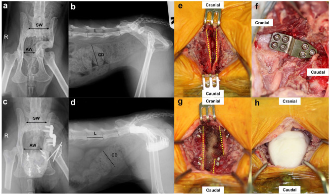
Objectives: The aim of this study was to assess the efficacy of pelvic osteotomy and ventral fixation of the ischium using cortical screws and polymethylmethacrylate (PMMA) for feline pelvic canal stenosis (PCS) associated with malunion after conservative management of pelvic fractures.
Methods: Surgical pelvic enlargement was performed for PCS in six cats. The medical records, including information on the patients, surgical procedures, defecation and complications, were reviewed. The sacral index (SI) and colonic:lumbar:vertebral ratio (CLVR) were evaluated based on pre- and postoperative radiographs.
Results: This study included five castrated male cats and one spayed female cat. Postoperative improvements in constipation and defecatory difficulty were noted in all cases. The postoperative SI was significantly higher (mean 0.93, range 0.72-1.13) than the preoperative SI (mean 0.59, range 0.45-0.74) (P <0.001). However, no statistically significant change was found in the CLVR preoperatively and up to 3 months postoperatively. A successful union of the ilium was observed, without implant failures. One case developed necrosis of the pubic surgical wound.
Conclusions and relevance: This study indicated the potential benefits of pelvic osteotomy and ventral fixation of the pelvic floor using screws and PMMA for achieving pelvic cavity enlargement in treating feline PCS associated with defecatory problems.
Keywords: Pelvic canal stenosis; pelvic osteotomy; polymethylmethacrylate; ventral fixation.
Conflict of interest statement
Conflict of interestHF is employed by Platon Japan, Tokyo, Japan, the manufacturer of crank plates and screws used in this study. The authors declared that there were no other conflicts of interest.
Figures

References
-
- Grierson J. Dealing with pelvic fractures in cats. In Practice 2019; 41: 106–114.
-
- Bookbinder PF, Flanders JA. Characteristics of pelvic fracture in the cat. Vet Comp Orthop Traumatol 1992; 5: 122–127.
-
- Hamilton MH, Evans DA, Langley-Hobbs SJ. Feline iliac fractures: assessment of screw loosening and pelvic canal narrowing after lateral plating. Vet Surg 2009; 38: 326–333. - PubMed
-
- Colopy-Poulsen SA, Danova NA, Hardie RJ, et al. Managing feline obstipation secondary to pelvic fracture. Comp Contin Educ Pract Vet 2005; 27: 662–669.
MeSH terms
LinkOut - more resources
Full Text Sources
Miscellaneous
