. 2025 Jan;24(1):e14362.
doi: 10.1111/acel.14362. Epub 2024 Oct 31.

Morphological phenotyping of the aging cochlea in inbred C57BL/6N and outbred CD1 mouse strains

Affiliations

Morphological phenotyping of the aging cochlea in inbred C57BL/6N and outbred CD1 mouse strains

Chiara Attanasio et al. Aging Cell. 2025 Jan.

Abstract

Morphological mouse phenotyping plays a pivotal role in the translational setting and even more in the area of auditory research, where mouse is a central model organism due to the evolutionary genetic relationship and morpho-functional analogies with the human auditory system. However, some results obtained in murine models cannot be translated to humans due to the inadequate description of experimental conditions underlying poor reproducibility. We approach the characterization of the aging process of the mouse cochlea in animals up to 18 months of age belonging to two of the most used outbred (CD1) and inbred (C57BL/6N) strains. Striving to reduce any environmental variable we performed our study compliantly to the ARRIVE guidelines. We integrated instrumental data (auditory brainstem response test), with morphological analyses to correlate functional discrepancies to morphological changes and track the differences in the evolution of sensorineural hearing loss in the two strains. We featured the localization of Gipc3, Myosin VIIa, and TMC1 in hair cells of the Corti organ as well as NF 200 and the density of type I neuron in the spiral ganglion. We outlined age-related hearing loss (ARHL) in both strains, and a clear drop in the selected marker localization. However, in CD1 we detected a different trend allowing the identification of potential strain-specific mechanisms, namely an increase in myosin VIIa in 6 months aging mice in comparison to 2 months old animals. Our findings represent an asset to investigate the strain-dependent physiological trigger of ARHL providing new insights in the translational area.

Keywords: aging cochlea; animal models; morphological phenotyping; mouse strains; sensorineural hearing loss.


Conflict of interest statement

All authors disclosed no conflicts of interest.

Figures
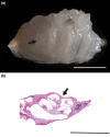
FIGURE 1
FIGURE 1
Dissected Cochlea: (a) shows the appearance of the dissected mouse cochlea, (b) displays the histological section of the tissue stained by Hematoxylin/Eosin (H‐E). Arrows indicate the modiulus. Scale bars 600 μm.
FIGURE 2
FIGURE 2
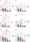
ABR results: The diagrams show the results of auditory brainstem response test in CD1 and C57BL/6N mice in both sexes at different ages: (a) 2 months, (b) 6 months, (c) 12 months, and (d) 18 months. Within the same diagram the results are grouped by type and frequencies of the acoustic stimuli (click threshold, 8000 Hz, 16,000 Hz and 24,000 Hz). Differences between groups were analyzed by ANOVA followed by Tukey's HSD post‐hoc test. p values <0.05 (*), p values <0.01 (**), and p values <0.0001 (****) have been considered statistically significant.
FIGURE 3
FIGURE 3
Hair cells specific markers: Immunofluorescence to Gipc3, MyoVIIa, and TMC1 in the organ of Corti of C57BL/6N mice at 2, 6, 12 and 18 months of age The images are representative of both the strains. Red arrows indicate inner hair cells in each section. Scale bars 20 μm.
FIGURE 4
FIGURE 4
Hair cell markers quantitative image analysis: The histograms display the quantitative analysis of the immunofluorescence toGipc3, MyoVIIa, and TMC1 in hair cells. (a, a1, a2) Comparisons between the different timepoints, 2, 6, 12 and 18 months for each strain. (b, b1, b2) Comparisons between the two strains, C57BL/6N versus CD1, at each timepoint. Differences between groups were analyzed by ANOVA followed by Tukey's HSD post‐hoc test. p values <0.05 (*), p values <0.01 (**), p values <0.001 (***), and p values <0.0001 (****) have been considered statistically significant.
FIGURE 5
FIGURE 5
Spiral ganglion and neuronal density: (a) Hematoxylin/Eosin (H–E) staining of spiral ganglia of C57BL/6N and CD1 mice at 2, 6, 12, and 18 months of age. Scale bars 100 μm. (b, c) The histograms display the density of spiral ganglion neurons. (b) Comparisons between the different timepoints, 2, 6, 12 and 18 months for each strain. (c) Comparisons between the two strains, C57BL/6N versus CD1, at each timepoint. Data were analyzed by ANOVA followed by Tukey's HSD post‐hoc test. p values <0.01 (**), p values <0.001 (***), and p values <0.0001 (****) were considered statistically significant.
FIGURE 6
FIGURE 6
NF 200 in spiral ganglion neurons and quantitative analysis: (a1, a2) Immunofluorescence to NF200 in the spiral ganglia of C57BL/6N mice at 2, 6, 12, and 18 months of age at low (a1) and high (a2) magnification. The images are representative of both the strains. Scale bars 20 μm. (b, c) The histograms display the quantitative results of spiral ganglion neuron counts expressed as relative density of NF200 positive cells in the spiral ganglion area. (b) Comparisons between the different timepoints, 2, 6, 12 and 18 months for each strain. (c) Comparisons between the two strains, C57BL/6N versus CD1, at each timepoint. Differences between groups were analyzed by ANOVA followed by Tukey's HSD post‐hoc test. p values <0.05 (*), p values <0.01 (**), and p values <0.0001 (****) were considered statistically significant.

References

    1. Biggers, J. D. , & Claringbold, P. J. (1954). Why use inbred lines? Nature, 174(4430), 596–597. 10.1038/174596a0 - DOI - PubMed
    1. Bowl, M. R. , & Dawson, S. J. (2015). The mouse as a model for age‐related hearing loss—A mini‐review. Gerontology, 61(2), 149–157. 10.1159/000368399 - DOI - PubMed
    1. Carricondo, F. , & Romero‐Gómez, B. (2019). The Cochlear spiral ganglion neurons: The auditory portion of the VIII nerve. Anatomical Record (Hoboken, N.J.: 2007), 302(3), 463–471. 10.1002/ar.23815 - DOI - PubMed
    1. Castaño‐González, K. , Köppl, C. , & Pyott, S. J. (2024). The crucial role of diverse animal models to investigate cochlear aging and hearing loss. Hearing Research, 445, 108989. 10.1016/j.heares.2024.108989 - DOI - PubMed
    1. Charizopoulou, N. , Lelli, A. , Schraders, M. , Ray, K. , Hildebrand, M. S. , Ramesh, A. , Srisailapathy, C. R. S. , Oostrik, J. , Admiraal, R. J. C. , Neely, H. R. , Latoche, J. R. , Smith, R. J. H. , Northup, J. K. , Kremer, H. , Holt, J. R. , & Noben‐Trauth, K. (2011). Gipc3 mutations associated with audiogenic seizures and sensorineural hearing loss in mouse and human. Nature Communications, 2, 201. 10.1038/ncomms1200 - DOI - PMC - PubMed

LinkOut - more resources