. 2022 May 23;3(2):300-310.
doi: 10.3390/neurosci3020021. eCollection 2022 Jun.

Long COVID and the Autonomic Nervous System: The Journey from Dysautonomia to Therapeutic Neuro-Modulation through the Retrospective Analysis of 152 Patients

Affiliations

Long COVID and the Autonomic Nervous System: The Journey from Dysautonomia to Therapeutic Neuro-Modulation through the Retrospective Analysis of 152 Patients

Joseph Colombo et al. NeuroSci. .

Abstract

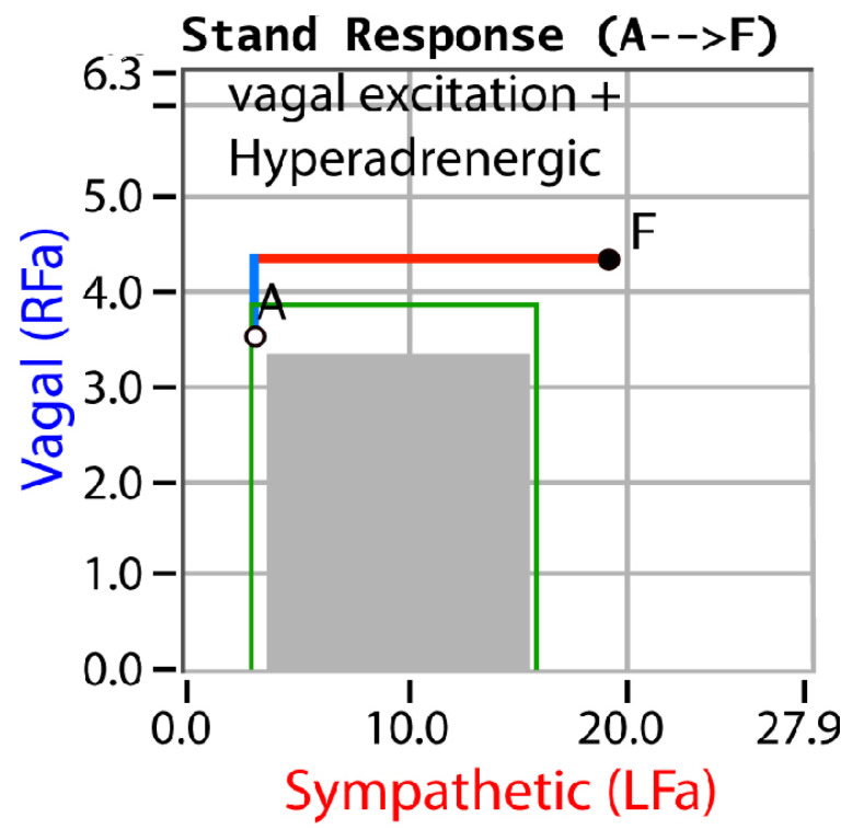
Introduction: The severity and prevalence of Post-Acute COVID-19 Sequela (PACS) or long-COVID syndrome (long COVID) should not be a surprise. Long-COVID symptoms may be explained by oxidative stress and parasympathetic and sympathetic (P&S) dysfunction. This is a retrospective, hypothesis generating, outcomes study.

Methods: From two suburban practices in northeastern United States, 152 long COVID patients were exposed to the following practices: (1) first, they were P&S tested (P&S Monitor 4.0; Physio PS, Inc., Atlanta, GA, USA) prior to being infected with COVID-19 due to other causes of autonomic dysfunction; (2) received a pre-COVID-19 follow-up P&S test after autonomic therapy; (3) then, they were infected with COVID-19; (4) P&S tested within three months of surviving the COVID-19 infection with long-COVID symptoms; and, finally, (5) post-COVID-19, follow-up P&S tested, again, after autonomic therapy. All the patients completed autonomic questionnaires with each test. This cohort included 88 females (57.8%), with an average age of 47.0 years (ranging from 14 to 79 years), and an average BMI of 26.9 #/in2.

Results: More pre-COVID-19 patients presented with sympathetic withdrawal than parasympathetic excess. Post-COVID-19, these patients presented with this ratio reversed and, on average, 49.9% more autonomic symptoms than they did pre-COVID-19.

Discussion: Both parasympathetic excess and sympathetic withdrawal are separate and treatable autonomic dysfunctions and autonomic treatment significantly reduces the prevalence of autonomic symptoms.

Conclusion: SARS-CoV-2, via its oxidative stress, can lead to P&S dysfunction, which, in turn, affects the control and coordination of all systems throughout the whole body and may explain all of the symptoms of long-COVID syndrome. Autonomic therapy leads to positive outcomes and patient quality of life may be restored.

Keywords: autonomic dysfunction; autonomic therapy; long COVID; outcomes; parasympathetic; sympathetic.


Conflict of interest statement

Conflicts of InterestOnly Colombo has a conflict of interest as Co-Founder and Sr. Medical Director of Physio PS, Inc. No other individual associated with this manuscript has a conflict of interest.

Figures

Figure 1
Figure 1
“Normal at Rest”. A resting (baseline) P&S response plot depicting normal and abnormal ranges. The gray area depicts the normal response region. The purple highlighted areas depict the definitions of Advanced Autonomic Dysfunction (AAD, light purple) or Diabetic Autonomic Neuropathy (DAN, also light purple), and Cardiovascular Autonomic Neuropathy (CAN, dark purple). AAD and DAN indicate an increased morbidity risk and CAN indicates an increased mortality risk. Risk is stratified by sympathovagal balance (“LFa/RFa” = S/P). The space between the two outer diagonal lines defines a normal sympathovagal balance, regardless of the resting autonomic state. A normal sympathovagal balance normalizes the morbidity and mortality risks. Above and to the left of the upper diagonal line indicates a low sympathovagal balance, which is a resting parasympathetic excess. Below and to the right of the lower diagonal line indicate a high sympathovagal balance, which is a resting sympathetic excess.
Figure 2
Figure 2
“Normal upon Standing”. An example normal stand P&S response plot. Active standing is equivalent to a positive, head-up, tilt [25]. Point “A” is the patient’s resting, baseline response and point “F” is the patient’s stand response. In the normal stand response, the parasympathetics (the blue line) first decrease and then the (alpha-)sympathetics (the red line) increase [5].
Figure 3
Figure 3
“Sympathetic Withdrawal”. An example of an abnormal stand P&S response plot depicting alpha-sympathetic withdrawal. Point “A” is the patient’s resting, baseline response and point “F” is the patient’s stand response. Here, the parasympathetic response is normal (see Figure 2), but the sympathetic response decreases abnormally, indicating orthostatic dysfunction, possibly leading to all forms of POTS, orthostatic intolerance, orthostatic hypotension, neurogenic orthostatic hypotension, etc. [5].
Figure 4
Figure 4
“Vagal Excitation”. An example of an abnormal stand P&S response plot depicting parasympathetic excess. Point “A” is the patient’s resting, baseline response and point “F” is the patient’s stand response. Here, the sympathetic response is normal (see Figure 2), but the parasympathetic response increases abnormally, indicating vagal or parasympathetic excess, associated with difficult-to-control BP, blood glucose, hormone levels, or weight; difficult-to-describe pain syndromes (including CRPS); unexplained arrhythmia (palpitations) or seizures; temperature dysregulation (both the response to heat or cold and sweat responses); symptoms of depression or anxiety, ADD/ADHD, fatigue, exercise intolerance, sex dysfunction, sleep or GI disturbance, lightheadedness, cognitive dysfunction or “brain fog”; and frequent headaches or migraines. Parasympathetic excess and sympathetic withdrawal may concurrently occur, including the fact that parasympathetic excess may mask sympathetic withdrawal. This masking is indicated by an abnormal BP response to stand as compared with resting BP [5].
Figure 5
Figure 5
“Vagal Excitation + Hyperadrenergic”. An example of an abnormal stand P&S response plot depicting parasympathetic excess with sympathetic excess. Point “A” is the patient’s resting, baseline response and point “F” is the patient’s stand response. Here, the parasympathetic response is abnormal (see Figure 2), as is the sympathetic response which increases too significantly, exceeding the normal area. The combination indicates vasovagal syncope. The parasympathetic excess is the vagal component, and the sympathetic excess (hyperadrenergic response) indicates the nervous system’s response to syncope and the accompanying poor cerebral perfusion [5].

Similar articles

Cited by

References

    1. DePace N.L., Colombo J. Long-covid syndrome: A multi-organ disorder. Cardio. Open. 2022;7:212–223.
    1. Alwan N.A., Johnson L. Defining long COVID: Going back to the start. Med. 2021;2:501–504. doi: 10.1016/j.medj.2021.03.003. - DOI - PMC - PubMed
    1. DePace N.L., Colombo J. Long-COVID syndrome: A review of what we have learned clinically to date. 2022. Submitted .
    1. DePace N.L., Colombo J. Autonomic and Mitochondrial Dysfunction in Clinical Diseases: Diagnostic, Prevention, and Therapy. Springer Science + Business Media; New York, NY, USA: 2019.
    1. Colombo J., Arora R.R., DePace N.L., Vinik A.I. Clinical Autonomic Dysfunction: Measurement, Indications, Therapies, and Outcomes. Springer Science + Business Media; New York, NY, USA: 2014.

LinkOut - more resources