Case Report: Does the misplaced titanium mesh cage after total spondylectomy causing cervicothoracic cord compression need to be removed during revision surgery?
- PMID: 39483375
- PMCID: PMC11524943
- DOI: 10.3389/fsurg.2024.1394135
Case Report: Does the misplaced titanium mesh cage after total spondylectomy causing cervicothoracic cord compression need to be removed during revision surgery?
Abstract
Background: Mechanical failure following total spondylectomy is a surgical challenge. The cervicothoracic junction region is a special anatomical site with complex biomechanics, and few studies have reported a detailed surgical management strategy for cases where the mesh cage subsides and compresses the spinal cord in the cervicothoracic junction region after total spondylectomy.
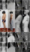
Case presentation: A 56-year-old male patient experienced screw and rod fracture and mesh cage retropulsion into the spinal canal 5 years after total spondylectomy for osteochondroma in the first to third thoracic vertebrae. The patient complained of numbness and discomfort in both lower extremities, accompanied by unstable walking for 8 months prior to admission at our hospital. We concluded that uncorrected local kyphosis in the cervicothoracic junction after the first surgery resulted in current mesh cage subsidence and rod/screw fracture. Considering the difficulty and risks of removing the mesh cage from the anterior approach, we initially freed the superior end of the mesh cage without removing the mesh from the anterior approach by resecting the C6/7 intervertebral disc and the destroyed C7 vertebral body. We then removed the original screws and rods and performed long segment fixation from C4 to T6 via a posterior approach after recovering sagittal alignment by skull traction. Finally, the iliac bone was harvested and transplanted between the superior end of the mesh cage and the inferior end plate of C6 to fill the defect caused by kyphosis correction and C7 vertebral resection. After surgery, the patient experienced sagittal alignment reconstruction and symptom relief, and he was asked to wear a cast for at least 6 months until bone fusion was achieved. At the 3-year follow-up, there was fusion between the mesh cage and the C6 vertebra with successful instrument reconstruction and no mesh cage subsidence were observed.
Conclusions: When a subsided and migrated titanium mesh cage is difficult to remove after mechanical failure following total spondylectomy, recovering sagittal alignment to achieve indirect decompression based on unique anterior and middle column reconstruction, solid instrument construction, and bone fusion is an alternative solution.
Keywords: cage subsidence; cervicothoracic junction; mechanical failure; revision surgery; total spondylectomy.
© 2024 Wang, Cheng, Zhao and Zhao.
Conflict of interest statement
The authors declare that the research was conducted in the absence of any commercial or financial relationships that could be construed as a potential conflict of interest.
Figures


References
-
- Salame K, Regev G, Keynan O, Lidar Z. Total en bloc spondylectomy for vertebral tumors. Isr Med Assoc J IMAJ. (2015) 17:37–41. - PubMed
Publication types
LinkOut - more resources
Full Text Sources
Miscellaneous

