. 2022 Oct 23;3(4):566-588.
doi: 10.3390/neurosci3040041. eCollection 2022 Dec.

The Effect of Doxapram on Proprioceptive Neurons: Invertebrate Model

Affiliations

The Effect of Doxapram on Proprioceptive Neurons: Invertebrate Model

Bethany J Ison et al. NeuroSci. .

Abstract

The resting membrane potential enables neurons to rapidly initiate and conduct electrical signals. K2p channels are key in maintaining this membrane potential and electrical excitability. They direct the resting membrane potential toward the K+ equilibrium potential. Doxapram is a known blocker for a subset of K2p channels that are pH sensitive. We assessed the effects of 0.1 and 5 mM doxapram on the neural activity within the propodite-dactylopodite (PD) proprioceptive sensory organ in the walking legs of blue crabs (Callinectes sapidus). Results indicate that 0.1 mM doxapram enhances excitation, while the higher concentration 5 mM may over-excite the neurons and promote a sustained absolute refractory period until the compound is removed. The effect of 5 mM doxapram mimics the effect of 40 mM K+ exposure. Verapamil, another known K2p channel blocker as well as an L-type Ca2+ channel blocker, reduces neural activity at both 0.1 and 5 mM. Verapamil may block stretch activated channels in sensory endings, in addition to reducing the amplitude of the compound action potential with whole nerve preparations. These findings are notable as they demonstrate that doxapram has acute effects on neurons of crustaceans, suggesting a targeted K2p channel. The actions of verapamil are complex due to the potential of affecting multiple ion channels in this preparation. Crustacean neurons can aid in understanding the mechanisms of action of various pharmacological agents as more information is gained.

Keywords: K2p channels; crab; proprioception; sensory.


Conflict of interest statement

Conflicts of InterestThe authors declare that they have no conflict of interest.

Figures

Figure A1
Figure A1
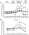
The activity obtained from the PD nerve for five different groups within a university course for the effect of doxapram (5 mM). (A) The number of spikes were measured in the same way and by the same person who measured all the traces independently of the student participants for consistency in analysis. (B) The number of spikes in each of the three trials was averaged and graphed in the same manner as in (A), which allows an easier view of the overall effects.
Figure A2
Figure A2
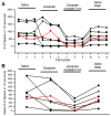
Four groups of different participants, with two people per group, analyzed the same data sets (A #1 and B #2). (A) The four groups who analyzed the #1 data set fits well with the analysis from a master in analysis, whose counts are indicated by the red line. The master analyzed all data sets in the study. (B) The participants deviated from the master in analysis for three of the four groups in the condition where the doxapram had been incubated for 5 min. The other experimental conditions followed a similar trend as that of the master.
Figure A3
Figure A3
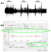
Analysis of data with the same automated measure. Two traces are provided: three trials in saline (A1) and the three trials in doxapram after 5 min of incubation (B1). The automated analysis is set to 2 SD (standard deviations of the mean in the traces; see small green ellipse in (A2,B2)) for both (A1,B1), but it produces differences in what is detected, as indicated in the large green ellipse in (A2,B2). The open circles inside the ellipse indicate what deflections in the trace are counted as spikes. Since there is little neural activity in (B1), the noise of the trace is being detected as spikes, producing an artificially high number of spikes.
Figure A3
Figure A3
Analysis of data with the same automated measure. Two traces are provided: three trials in saline (A1) and the three trials in doxapram after 5 min of incubation (B1). The automated analysis is set to 2 SD (standard deviations of the mean in the traces; see small green ellipse in (A2,B2)) for both (A1,B1), but it produces differences in what is detected, as indicated in the large green ellipse in (A2,B2). The open circles inside the ellipse indicate what deflections in the trace are counted as spikes. Since there is little neural activity in (B1), the noise of the trace is being detected as spikes, producing an artificially high number of spikes.
Figure 1
Figure 1
The first or second walking leg of the crab was used to expose the PD organ and associated nerve to various compounds. The joint was initially bent at 90 degrees, then extended out straight within 1 s, and then held for at least another 9 s. The entire 10 s was then used for analysis in the number of spikes that occurred while bathed in different solutions.
Figure 2
Figure 2
Representation of the effects of doxapram at 5 mM on neural activity for the proprioceptive neurons in the crab PD organ. (A) The activity of the nerve in saline with the three movements of the joint (1 s for the movement to an extended position and 9 s or more for being held in a static position of joint extension). (B) After the bath is exchanged to doxapram, the joint is then moved again three times. (C) Flushing the doxapram solution around the preparation and allowing it to incubate for 5 min. After 5 min, three more movements are made. (D) The bath is exchanged two times with fresh saline and the joint movements are repeated. Only the initial 10 s are used for analysis.
Figure 3
Figure 3
The acute effect of doxapram (5 mM) on neural activity of the PD organ. (A) The number of spikes measured in the 10-s window from the beginning movement of the joint starting from a bent position (90 degrees) to fully extended within 1 s and held in an extended position for the next 9 s. This paradigm is repeated three times for each condition. Each line represents a different preparation of an PD organ. Three trials were undertaken with saline, three trials were done immediately after switching the bath to doxapram (5 mM), and they were examined again after incubation for 5 min. The final exchange was to rinse the preparation twice with fresh saline and then move the joint three more times. Each movement was separated by at least 10 s while the joint was held in a bent position. (B) The number of spikes in each of the three trails was averaged and graphed in the same manner as in (A), which allows an easier view of the overall effects. The red colored trace represents a PD preparation from a chela of the large claw.
Figure 4
Figure 4
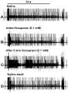
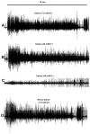
Representative responses to the effects of 0.1 doxapram exposure. The second trial of the three movements for each condition: (A) initial saline, (B) initial exposure to doxapram, (C) 5 min of incubation to doxapram, and (D) saline wash out. The number of spikes within the initial 10 s is used for quantification.
Figure 5
Figure 5
The acute effect of doxapram (0.1 mM) on neural activity of the PD organ. (A) The number of spikes measured in the 10 s window from the beginning movement of the joint starting from a bent position (90 degrees) to fully extended within 1 s and held in an extended position for the next 9 s. This paradigm is repeated three times for each condition. Each line represents a different preparation of a PD organ. Three trials were undertaken with saline, three trials were done immediately after switching the bath to doxapram (0.1 mM), and they were examined again after incubation for 5 min. The final exchange was to rinse the preparation twice with fresh saline and then move the joint three more times. Each movement was separated by at least 10 s while the joint was held in a bent position. (B) The number of spikes in each of the three trials was averaged and graphed in the same manner as in (A), which allows an easier view of the overall effects.
Figure 6
Figure 6
The effect of raised extracellular K+ on the neural activity for a representative PD organ. The activity of the PD nerve over 10 s when the joint from a 90-degree angle is fully extended within 1 sec and held for 9 s. The activity in saline (A) to saline containing 20 mM K+ (B). The 20 mM K+ did not show a significant change in overall activity. A change to a saline with 40 mM K+ (C) decreased the activity substantially. Some activity is regained with bathing the preparation in fresh saline (D).
Figure 7
Figure 7
The number of spikes within 10 s when displacing the joint and holding it in a static position for six different preparations. There is no a significant effect for exposure to 20 mM K+ but there is a significant decrease in activity when exposed to 40 mM K+ (p < 0.05 paired T-Test).
Figure 8
Figure 8
A representative effect of verapamil (5 mM) on neural activity of the PD nerve during the extension and bending of the PD joint. The movement of the joint is shown at the top and the 10 s used for analysis in the number of spikes recorded are shown for each paradigm. Three trials of movement are used initially during saline and when switching the bath to verapamil and following the flushing of the recording chamber with fresh saline.
Figure 9
Figure 9
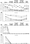
The acute effect of verapamil (0.1 mM and 5 mM) on neural activity of the PD organ. (A1) The number of spikes measured in the 10 s window from the beginning movement of the joint starting from a bent position (90 degrees) to fully extended within 1 s and held in an extended position for the next 9 s. This paradigm is repeated three times for each condition. Each line represents a different preparation of a PD organ. Three trials were undertaken with saline, three trials were done immediately after switching the bath to verapamil (0.1 mM), and they were examined again after incubation for 5 and 20 min. The last step was to rinse the preparation with fresh saline twice and then move the joint three more times. Each movement was separated by at least 10 s while the joint was held in a bent position. (A2) The number of spikes in each of the three trials was averaged and graphed in the same manner as in (A1), which allows an easier view of the overall effects. (B1) The same analysis is shown for the three trials in each condition for exposure to 5 mM verapamil. (B2) The average of each of the three trials for each condition is shown. Depression in neural activity was present at 0.1 and 5 mM after 20 min (p < 0.05; paired T-Test; N = 6 for each concentration). However, activity was depressed even after the initial exposure to 5 mm (p < 0.05; paired T-Test; N = 6).
Figure 10
Figure 10
Representative effects of verapamil on the compound action potentials (CAPs) of the walking leg nerve. The CAPs in saline (A1,B1) and after 20 min of exposure to 0.1 mM (A2) or 5 mM (B2) of verapamil showed some depression for both concentrations. (A3,B3) Removal of verapamil with fresh saline did not fully recover the amplitude of the CAPs.
Figure 11
Figure 11
The percent change in the areas of the trace for the compound action potentials (CAPs) in saline and after 20 min of exposure to 0.1 mM or 5 mM of verapamil. The percent change in the area of the CAPs from initial saline to the wash-out is also shown. Some depression still occurred even after washout for both concentrations. (p < 0.05; paired T-Test; N = 6 for each concentration from initial saline to after 20 min of exposure to verapamil).
Figure 12
Figure 12
A representative model to explain the observed phenomenon with exposure to doxapram in relation to the neural activity in sensory neurons of the PD organ in a crab preparation. (A) A representative sensory neuron which is activated by opening stretch activated ion channels (SACs). The depolarization from activating SACs may reach the threshold to open voltage-gated Na+ channels (Nav+) and, subsequently, voltage-gated K+ channels (Kv+) to allow action potentials to travel along the nerve (see inset in top right corner). The K2p channels help to maintain the resting membrane potential along with the Na+-K+ ATP dependent pump. (B) In the presence of doxapram, the K2p channels are blocked. (C) The effect of blocking the K2p channels depolarizes the neurons. A low level may bring the membrane closer to threshold to activate the Nav+ channels and produce more action potentials for the same stimulus. However, if the neuron depolarizes and cannot repolarize, the inactivation of the Nav+ channels would result in prolonged absolute refractory periods while exposed to doxapram, not allowing the neuron to be excitable. (D) The potential effects of verapamil are illustrated. The SACs are likely blocked rapidly upon exposure to low and high concentrations. On the axon, the voltage gated Ca2+ channels (Cav2+) as well as the Nav+ may be a target given the compound action potential is slowly depressed over time, independent of the actions on the SACs in the sensory endings.

References

    1. Buckingham S.D., Kidd J.F., Law R.J., Franks C.J., Sattelle D.B. Structure and function of two-pore-domain K+ channels: Contributions from genetic model organisms. Trends Pharmacol. Sci. 2005;26:361–367. doi: 10.1016/j.tips.2005.05.003. - DOI - PubMed
    1. Goldstein S.A., Price L.A., Rosenthal D.N., Pausch M.H. ORK1, a potassium-selective leak channel with two pore domains cloned from Drosophila melanogaster by expression in Saccharomyces cerevisiae. Proc. Natl. Acad. Sci. USA. 1996;93:13256–13261. doi: 10.1073/pnas.93.23.13256. - DOI - PMC - PubMed
    1. Goldstein S.A., Wang K.W., Ilan N., Pausch M.H. Sequence and function of the two P domain potassium channels: Implications of an emerging superfamily. J. Mol. Med. 1998;76:13–20. doi: 10.1007/s109-1998-8100-0. - DOI - PubMed
    1. Plant L.D., Goldstein S.A.N. Two-Pore Domain Potassium Channels. In: Zheng J., Trudeau M.C., editors. Handbook of Ion Channels. 1st ed. CRC Press; Boca Raton, FL, USA: 2015.
    1. Enyedi P., Czirják G. Molecular background of leak K+ currents: Two-pore domain potassium channels. Physiol. Rev. 2010;90:559–605. doi: 10.1152/physrev.00029.2009. - DOI - PubMed

LinkOut - more resources