[Preprint]. 2024 Oct 16:rs.3.rs-5263500.
doi: 10.21203/rs.3.rs-5263500/v1.

Targeting the Galectin-1/Ras Interaction for Treating Malignant Peripheral Nerve Sheath Tumors

Affiliations

Targeting the Galectin-1/Ras Interaction for Treating Malignant Peripheral Nerve Sheath Tumors

Hsiao-Chi Wang et al. Res Sq. .

Abstract

Background: Neurofibromatosis type 1 (NF1) is a common inherited neurological disorder that can lead to the development of malignant peripheral nerve sheath tumors (MPNSTs), a highly aggressive form of sarcoma. Current treatment options for MPNSTs are limited, with poor prognosis and high recurrence rates. This study aims to explore the potential of targeting the Galectin-1 (Gal-1) and Ras interaction as a novel therapeutic strategy for MPNSTs.

Methods: Molecular docking simulations were conducted to identify specific residues involved in the Gal-1 and H-Ras(G12V) interaction. LLS30, a compound designed to target the Ras binding pocket on Gal-1, was developed and tested. The efficacy of LLS30 was evaluated through in vitro assays, including cell viability, apoptosis, and co-immunoprecipitation studies, as well as in vivo assays using orthotopic MPNST xenograft and experimental lung metastasis models. Transcriptome sequencing was performed to analyze the impact of LLS30 on gene expression and signaling pathways.

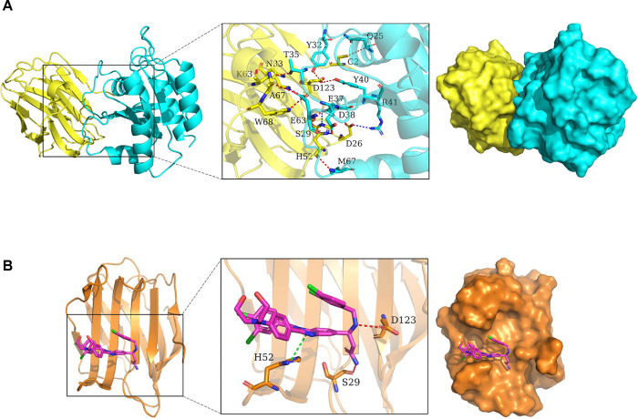
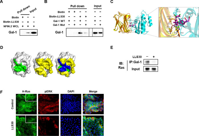
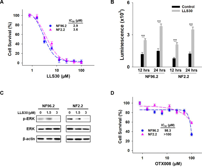
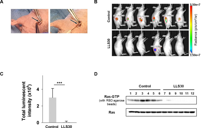
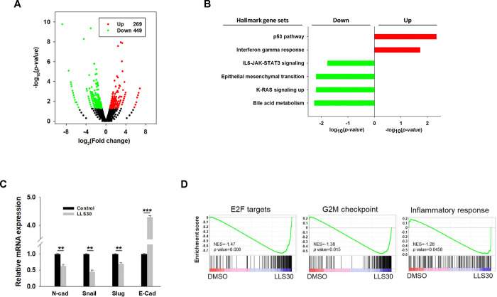
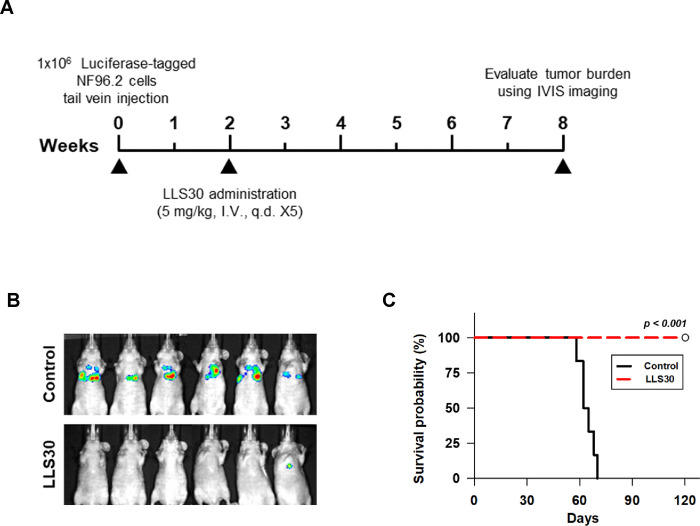
Results: Molecular docking revealed key residues involved in the Gal-1/Ras interaction, and LLS30 was shown to bind to these residues, disrupting the interaction. LLS30 treatment resulted in Ras delocalization from the plasma membrane and suppression of the Ras/Erk signaling pathway. In vitro, LLS30 significantly reduced MPNST cell proliferation and induced apoptosis. In vivo, LLS30 demonstrated potent anti-tumor activity, reducing tumor burden and metastasis while improving survival in animal models. Transcriptome analysis showed that LLS30 downregulates critical pathways, including KRAS signaling and epithelial-mesenchymal transition (EMT).

Conclusions: Interference with the Gal-1/Ras interaction could lead to suppression of the Ras signaling pathway. LLS30 effectively disrupts the Gal-1/Ras interaction, resulting in significant anti-tumor and anti-metastatic effects in MPNST models. These findings indicated that targeting Gal-1 with LLS30 offers a promising therapeutic approach for treating MPNSTs and may also be applicable to other malignancies where Gal-1 and Ras are key oncogenic drivers.


Conflict of interest statement

Conflict of Interest Author Hsiao-Chi Wang and Tsung-Chieh Shih are employed by Kibio Inc. The remaining authors declare that the research was conducted in the absence of any commercial or financial relationships that could be construed as a potential conflict of interest. •Competing interests Author Hsiao-Chi Wang and Tsung-Chieh Shih are employed by Kibio Inc. The remaining authors declare that they have no competing interests.

Figures

Fig. 1.
Fig. 1.. Molecular dynamics simulation of Gal-1, Ras, and LLS30 interactions.
(A) The 3D binding model of Gal-1 with H-RAS(G12V). Gal-1 is colored with yellow, H-Ras(G12V) with cyan. The residues in Gal-1 are shown as yellow sticks, the residues in H-Ras(G12V) are shown as greencyan sticks. Red dashes indicate hydrogen bond interactions, while blue dashes represent salt bridges. (B) The 3D binding model of Gal-1 with LLS30. Gal-1 is colored with orange, LLS30 with magentas. The residues in Gal-1 are depicted as orange sticks. Red dashes represent hydrogen bond interaction while green dashes represent Pi-Pi conjugate.
Fig. 2.
Fig. 2.. LLS30 interacts with Gal-1 and disrupts Gal-1/Ras interactions in MPNST Cells.
(A) The pull-down assay confirmed the direct interaction between LLS30 and Gal-1 from NF96.2 whole cell lysate (WCL). Biotin-LLS30 conjugated with streptavidin-agarose beads was incubated with NF96.2 WCL to pull down interacting proteins. After thorough washing to remove non-specifically bound proteins, the proteins bound to the beads were eluted and confirmed by immunoblots using an antibody against Gal-1. The input lane represents NF96.2 WCL without pull-down, serving as an input control to show the baseline level of Gal-1 in the cells. (B) LLS30 pulls down Gal-1 wild type (WT) but exhibits reduced binding with Gal-1 mutant (D123A) (Mut). Biotin-LLS30 bound to streptavidin-agarose beads was incubated with recombinant proteins Gal-1 WT or Mut. After wash, the level of bound protein was detected via immunoblots with an anti-Gal-1 antibody. Gal-1 WT or Mut alone was loaded as input. (C) The superposition diagrams of the H-Ras(G12V)-Gal-1 complex and the Gal-1-LLS30 (magenta) complexes. (D) The binding region of LLS30 on the surface of the Gal-1 protein is shown in green (left), while the binding region of H-Ras(G12V) on the Gal-1 protein surface is depicted in yellow (middle); the overlapping region where LLS30 and H-Ras(G12V) bind on the Gal-1 protein surface is highlighted in blue (right). (E) Co-immunoprecipitation experiments investigated the interaction between Gal-1 and H-Ras after treatment with LLS30 or vehicle DMSO. F96.2 cells were first treated overnight with either LLS30 or DMSO. Subsequently, cell membrane proteins were extracted, followed by co-immunoprecipitation using an anti-Gal-1 antibody. The input consisted of total membrane protein extracted from NF96.2 cells without co-IP. Both the eluted proteins from co-IP and the total membrane protein without co-IP were analyzed by SDS-PAGE and immunoblotted with an anti-Ras antibody. (F) Immunofluorescence microscopy demonstrating the expression and localization of H-Ras and pERK in NF96.2 cells.
Fig 3.
Fig 3.. Effects of LLS30 in MPNST cells.
(A) Cell survival of NF96.2 and NF2.2 MPNST cells treated with indicated concentrations of LLS30 for 72 hours. IC50 of LLS30 on NF96.2 and NF2.2 are 2.9 μM and 3.6 μM, respectively. (B) Caspase-3/7 activities in NF96.2 and NF2.2 cells after 12 and 24 hours of treatment with 0.05% DMSO or 5μM LLS30. (C) Immunoblots of phospho-Erk, Erk and β-actin in NF96.2 and NF2.2 treated with 0, 1.5 or 3 μM LLS30 for 24 hours. (D) Effect of OTX008 on MPNST cell lines. Cell viability of NF96.2 and NF2.2 MPNST cells was assessed after treatment with indicated concentrations of OTX008 for 72 hours. ***p < 0.001; two-tailed Student’s t test. Data shown are mean ± s.d.
Fig 4.
Fig 4.. LLS30 suppresses MPNST growth in an orthotopic xenograft mouse model.
(A) Illustration of sciatic nerve retrieval for the injection of luciferase-tagged NF96.2 cells to establish the orthotopic xenograft mouse model. (B) Representative bioluminescent imaging at week 8 after LLS30 or vehicle treatment, and (C) quantification of tumor signals. (D) Assessment of activated Ras expression levels using a Ras-GTP pull-down assay and immunoblotted with anti-Ras antibody (upper panel). Total Ras expression in protein extracted from both LLS30-treated and vehicle-treated tumors was examined via immunoblot with anti-Ras antibody (lower panel). Samples in lanes 1–6 were treated with vehicle, while lanes 7–12 received LLS30 treatment. ***p < 0.001; two-tailed Student’s t test. Data shown are mean ± s.d.
Fig 5.
Fig 5.. Transcriptomic analysis for LLS30 effects in NF96.2 MPNST cells.
(A) Volcano plot. The log2 fold change (FC) and log10 p-value indicated the expression level and significance for each gene. Each dot represents one gene. Black dots represent no significant differential expressed genes between control and LLS30 treatment, the green dots represent downregulated genes (FC<1.5, p<0.05) and red dots represent upregulated genes (FC>1.5, p<0.05). (B) Hallmark gene set analysis of the upregulated genes (FC>1.5, p<0.05) and downregulated genes (FC<1.5, p<0.05) between control and LLS30 treatment. Pathways significantly enriched for upregulated genes are represented by red bars, while those enriched for downregulated genes are denoted by green bars. (C) qRT-PCR analysis of EMT markers, including N-cadherin, Snail, Slug, and E-cadherin, in NF96.2 cells treated with vehicle or LLS30 (5 μM) for 24 hours. (D) Gene set enrichment analysis (GSEA) of hallmark gene sets significantly enriched in LLS30 treated cells vs control, with NES indicating nominal enrichment score. A gene set shows significant enrichment at a p < 0.05. **p < 0.01, ***p < 0.001; two-tailed Student’s t test. Data shown are mean ± s.d.
Fig 6.
Fig 6.. NF96.2 lung metastases bearing mice were treated with LLS30.
(A) The timeline illustrates the LLS30 treatment protocol. (B) Bioluminescent images were captured for control (8.7% alcohol/8.7% Tween-80) or LLS30-treated mice at 8 weeks after the initial i.v. injection of luciferase-tagged NF96.2 cells. (C) The Kaplan-Meier plot depicts the survival of PBS (n = 6) and LLS30-treated mice (n = 6). ***p < 0.001; log-rank test.

Similar articles

References

    1. Evans DG, Howard E, Giblin C, Clancy T, Spencer H, Huson SM, et al. Birth incidence and prevalence of tumor-prone syndromes: estimates from a UK family genetic register service. American journal of medical genetics Part A. 2010;152a(2):327–32. - PubMed
    1. Jouhilahti E-M, Peltonen S, Callens T, Jokinen E, Heape AM, Messiaen L, et al. The development of cutaneous neurofibromas. Am J Pathol. 2011;178(2):500–5. - PMC - PubMed
    1. Gutmann DH, Ferner RE, Listernick RH, Korf BR, Wolters PL, Johnson KJ. Neurofibromatosis type 1. Nature Reviews Disease Primers. 2017;3(1):17004. - PubMed
    1. Bates JE, Peterson CR, Dhakal S, Giampoli EJ, Constine LS. Malignant peripheral nerve sheath tumors (MPNST): a SEER analysis of incidence across the age spectrum and therapeutic interventions in the pediatric population. Pediatric blood & cancer. 2014;61(11):1955–60. - PubMed
    1. Hirbe AC, Gutmann DH. The management of neurofibromatosis type 1-associated malignant peripheral nerve sheath tumors: challenges, progress, and future prospects. Expert Opinion on Orphan Drugs. 2017;5(8):623–31.

Publication types

LinkOut - more resources