. 2023 Oct 20;4(4):280-295.
doi: 10.3390/neurosci4040023. eCollection 2023 Dec.

The Effects of Lithium on Proprioceptive Sensory Function and Nerve Conduction

Affiliations

The Effects of Lithium on Proprioceptive Sensory Function and Nerve Conduction

Kaitlyn E Brock et al. NeuroSci. .

Abstract

Animals are exposed to lithium (Li+) in the natural environment as well as by contact with industrial sources and therapeutic treatments. Low levels of exposure over time and high volumes of acute levels can be harmful and even toxic. The following study examines the effect of high-volume acute levels of Li+ on sensory nerve function and nerve conduction. A proprioceptive nerve in the limbs of a marine crab (Callinectes sapidus) was used as a model to address the effects on stretch-activated channels (SACs) and evoked nerve conduction. The substitution of Li+ for Na+ in the bathing saline slowed nerve conduction rapidly; however, several minutes were required before the SACs in sensory endings were affected. The evoked compound action potential slowed in conduction and slightly decreased in amplitude, while the frequency of nerve activity with joint movement and chordotonal organ stretching significantly decreased. Both altered responses could be partially restored with the return of a Na+-containing saline. Long-term exposure to Li+ may alter the function of SACs in organisms related to proprioception and nerve conduction, but it remains to be investigated.

Keywords: conduction; crustacean; lithium; proprioception; recruitment; sensory.


Conflict of interest statement

Conflicts of InterestThe authors declare no conflict of interest.

Figures

Figure 1
Figure 1
The isolation of the PD nerve for electrophysiological recordings. (A) The leg segments are shown, and the chordotonal organs are named by the joint they monitor. (B) The PD organ spans the most distal joint in the limb between the propodite and dactylopodite. (C) The PD nerve branches away from the main leg nerve near the base of the chordotonal strand.
Figure 2
Figure 2
Exposing the PD nerve for recording nerve activity. (A) A length of PD nerve can be isolated from the main leg nerve and pulled into a suction electrode. The joint is bent (A) and extended (B) while nerve activity is recorded.
Figure 3
Figure 3
Set-up for recording activity of the PD nerve via compound action potentials (CAPs). The proximal end of the main leg nerve was used for the actual recording, while the distal end was used to provide stimulation to induce CAPs. In this arrangement, position-sensitive neurons are not firing since the sensory endings are removed.
Figure 4
Figure 4
The experimental paradigm behind joint displacement and spike analysis. The joint is displaced from a flexed position to an extended one across a single second, held in place for at least ten more, and then moved back to flexed position. This was repeated three times in each bathing solution. An average of the activity across all three trials was used in conjunction with the raw data to assess the effects of changing the medium. The duration of exposure for each preparation depended on the medium in question. The spike count was obtained by counting the number of spikes from the beginning of the movement (across one second) through the next nine seconds of static positioning, and it was used as an index of PD organ neural activity. The arrow marks the beginning of the movement.
Figure 5
Figure 5
The effects of acute Na+ replacement by Li+ on PD nerve activity during joint displacement and extension. (A) The number of spikes within three ten-second displacements in saline, after five minutes of exposure of LiCl (470 mM- replacement of NaCl), and during a saline wash-out. Individual preparations are indicated. (B) The averaged activity for the three trials in each condition. (Saline to incubation in Li+ after 5 min; n = 6; paired t-test; p > 0.05).
Figure 6
Figure 6
Representative activity of the PD nerve during joint displacement and extension for up to 30 min of Li+ substitution. (A) In saline with Na+. (B) After 15 min of incubation in saline with Na+ replaced by Li+. (C) After 30 min of incubation in saline with Na+ replaced by Li+. (D) After two wash-outs, flushes back to normal saline (containing Na+ and no Li+). Each trace is shown over a period of twelve seconds, illustrating the ten seconds from which analysis was conducted, one second of joint movement to an extended position, and nine more seconds of being held static before being moved back to the starting position. The arrows mark the beginning of each movement. Note that the activity prior to the joint movements also decreased with Li+ exposure.
Figure 7
Figure 7
The effects of acute Li+ replacement of Na+ on PD nerve activity during joint displacement and extension for up to 30 min. (A) The number of spikes observed during the three ten-second displacement trials in saline, after 15 and 30 min of exposure to Li+ (470 mM), and during a saline wash-out. Individual preparations are indicated. (B) The average activity across all three trials in each condition for each preparation. (There is a significant decrease in neuronal activity after 15 or 30 min of exposure to Li+; n = 6; paired t-test; * p > 0.05).
Figure 8
Figure 8
The effects of 30-min incubation time for the PD organ while exposed to saline only. (A) The number of spikes within the three, ten-second displacement trials in saline, after 15 and 30 min of exposure to saline only and during a saline wash-out. Individual preparations are indicated. (B) The average activity across the three trials in each condition for each preparation. There are no significant effects of saline exposure over time (i.e., 30 min) on neuronal activity from PD displacement (n = 6; paired t-test; p > 0.05).
Figure 9
Figure 9
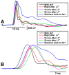
A representative preparation depicting the effects of Li+ replacement of Na+ on the leg nerve compound action potential (CAP) during evoked stimulation as superimposed traces. (A) Samples of CAPs before and during Li+ exposure at various times as well as during the return to normal saline with Na+, superimposed. (B) Enlarged images of superimposed traces are shown in A. Note: the conduction velocity slowed upon exposure to Li+ as well as the amplitude of the CAP peak.
Figure 10
Figure 10
The effects of acute Li+ replacement of Na+ on PD nerve activity during joint displacement and extension, taking place over a period of time up to 30 min and in seven different recording set-ups by seven different groups of researchers. (n = 6; * p < 0.05; paired t-test; initial saline to 15 min or to 30 min).

References

    1. Janka Z., Jones D.G. Lithium entry into neural cells via sodium channels: A morphometric approach. Neuroscience. 1982;7:2849–2857. doi: 10.1016/0306-4522(82)90107-5. - DOI - PubMed
    1. Obara S., Grundfest H. Effects of lithium on different membrane components of crayfish stretch receptor neurons. J. Gen. Physiol. 1968;51:635–654. doi: 10.1085/jgp.51.5.635. - DOI - PMC - PubMed
    1. Baldessarini R.J., Tondo L., Vázquez G.H. Pharmacological treatment of adult bipolar disorder. Mol. Psychiatry. 2019;24:198–217. doi: 10.1038/s41380-018-0044-2. - DOI - PubMed
    1. Fountoulakis K.N., Tohen M., Zarate C.A., Jr. Lithium treatment of Bipolar disorder in adults: A systematic review of randomized trials and meta-analyses. Eur. Neuropsychopharmacol. 2022;54:100–115. doi: 10.1016/j.euroneuro.2021.10.003. - DOI - PMC - PubMed
    1. Baird-Gunning J., Lea-Henry T., Hoegberg L.C.G., Gosselin S., Roberts D.M. Lithium poisoning. J. Intensive Care Med. 2017;32:249–263. doi: 10.1177/0885066616651582. - DOI - PubMed

LinkOut - more resources