

This is a preprint.
An open annotated dataset and baseline machine learning model for segmentation of vertebrae with metastatic bone lesions from CT
- PMID: 39484265
- PMCID: PMC11527073
- DOI: 10.1101/2024.10.14.24314447
An open annotated dataset and baseline machine learning model for segmentation of vertebrae with metastatic bone lesions from CT
Abstract
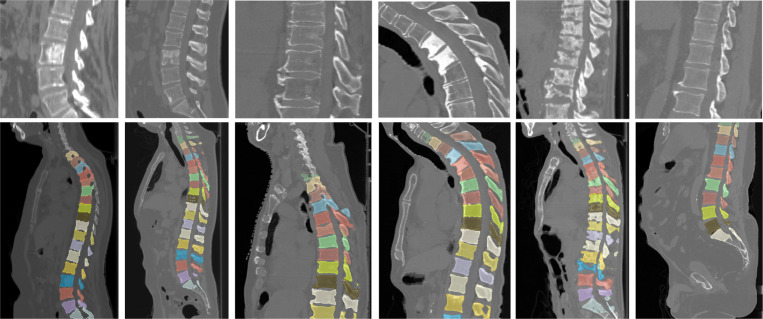
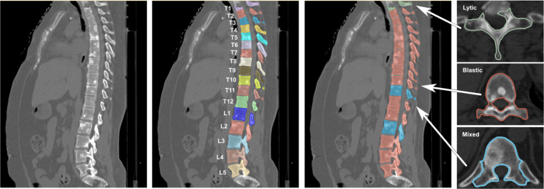
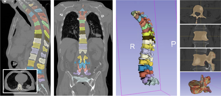
Automatic analysis of pathologic vertebrae from computed tomography (CT) scans could significantly improve the diagnostic management of patients with metastatic spine disease. We provide the first publicly available annotated imaging dataset of cancerous CT spines to help develop artificial intelligence frameworks for automatic vertebrae segmentation and classification. This collection contains a dataset of 55 CT scans collected on patients with various types of primary cancers at two different institutions. In addition to raw images, data include manual segmentations and contours, vertebral level labeling, vertebral lesion-type classifications, and patient demographic details. Our automated segmentation model uses nnU-Net, a freely available open-source framework for deep learning in healthcare imaging, and is made publicly available. This data will facilitate the development and validation of models for predicting the mechanical response to loading and the resulting risk of fractures and spinal deformity.
Figures
Similar articles
-
Segmentation of vestibular schwannoma from MRI, an open annotated dataset and baseline algorithm.Sci Data. 2021 Oct 28;8(1):286. doi: 10.1038/s41597-021-01064-w. Sci Data. 2021. PMID: 34711849 Free PMC article.
-
Can a Deep-learning Model for the Automated Detection of Vertebral Fractures Approach the Performance Level of Human Subspecialists?Clin Orthop Relat Res. 2021 Jul 1;479(7):1598-1612. doi: 10.1097/CORR.0000000000001685. Clin Orthop Relat Res. 2021. PMID: 33651768 Free PMC article.
-
Fully automatic segmentation of craniomaxillofacial CT scans for computer-assisted orthognathic surgery planning using the nnU-Net framework.Eur Radiol. 2022 Jun;32(6):3639-3648. doi: 10.1007/s00330-021-08455-y. Epub 2022 Jan 17. Eur Radiol. 2022. PMID: 35037088
-
The effect of deep learning-based lesion segmentation on failure load calculations of metastatic femurs using finite element analysis.Bone. 2024 Feb;179:116987. doi: 10.1016/j.bone.2023.116987. Epub 2023 Dec 5. Bone. 2024. PMID: 38061504
-
DentalSegmentator: Robust open source deep learning-based CT and CBCT image segmentation.J Dent. 2024 Aug;147:105130. doi: 10.1016/j.jdent.2024.105130. Epub 2024 Jun 13. J Dent. 2024. PMID: 38878813
References
-
- U.S. Cancer Statistics Working Group. U.S. Cancer Statistics Data Visualizations Tool bosd. Cancer Facts & Figures 2022. In: Institute CfDCaPaNC, editor.: U.S. Department of Health and Human Services; 2022.
-
- Siegel R, DeSantis C, Virgo K, Stein K, Mariotto A, Smith T, Cooper D, Gansler T, Lerro C, Fedewa S, Lin C, Leach C, Cannady RS, Cho H, Scoppa S, Hachey M, Kirch R, Jemal A, Ward E. Cancer treatment and survivorship statistics, 2012. CA Cancer J Clin. 2012;62(4):220–41. - PubMed
-
- Bilsky MH, Lis E, Raizer J, Lee H, Boland P. The diagnosis and treatment of metastatic spinal tumor. Oncologist 1999;4(6):459–69. - PubMed
-
- Sahgal A, Whyne CM, Ma L, Larson DA, Fehlings MG. Vertebral compression fracture after stereotactic body radiotherapy for spinal metastases. Lancet Oncol 2013;14(8):e310–20 - PubMed
-
- Rustoen T, Moum T, Padilla G, Paul S, Miaskowski C. Predictors of quality of life in oncology outpatients with pain from bone metastasis. J Pain Symptom Manage. 2005;30(3):234–42. - PubMed
Publication types
Grants and funding
LinkOut - more resources
Full Text Sources