[Preprint]. 2024 Oct 24:2024.10.23.619954.
doi: 10.1101/2024.10.23.619954.

The language network ages well: Preserved selectivity, lateralization, and within-network functional synchronization in older brains

Affiliations

The language network ages well: Preserved selectivity, lateralization, and within-network functional synchronization in older brains

Anne Billot et al. bioRxiv. .

Abstract

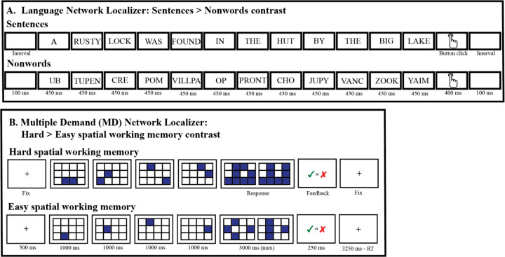
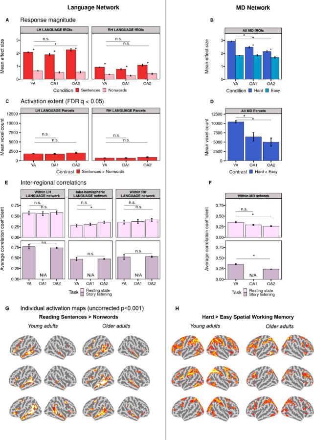
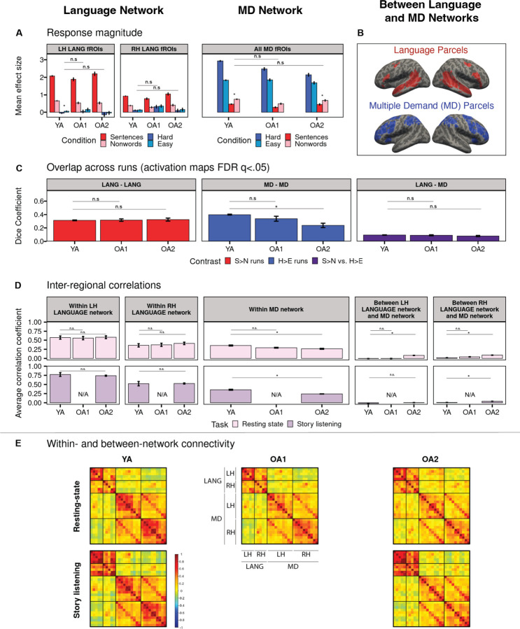
Healthy aging is associated with structural and functional brain changes. However, cognitive abilities differ from one another in how they change with age: whereas executive functions, like working memory, show age-related decline, aspects of linguistic processing remain relatively preserved (Hartshorne et al., 2015). This heterogeneity of the cognitive-behavioral landscape in aging predicts differences among brain networks in whether and how they should change with age. To evaluate this prediction, we used individual-subject fMRI analyses ('precision fMRI') to examine the language-selective network (Fedorenko et al., 2024) and the Multiple Demand (MD) network, which supports executive functions (Duncan et al., 2020), in older adults (n=77) relative to young controls (n=470). In line with past claims, relative to young adults, the MD network of older adults shows weaker and less spatially extensive activations during an executive function task and reduced within-network functional synchronization. However, in stark contrast to the MD network, we find remarkable preservation of the language network in older adults. Their language network responds to language as strongly and selectively as in younger adults, and is similarly lateralized and internally synchronized. In other words, the language network of older adults looks indistinguishable from that of younger adults. Our findings align with behavioral preservation of language skills in aging and suggest that some networks remain young-like, at least on standard measures of function and connectivity.

Keywords: aging; functional connectivity; functional localization; language network; lateralization; multiple demand network.


Conflict of interest statement

Competing Interest Statement: Dr. Kiran is a scientific advisor for Constant Therapy Health, but there is no overlap between this role and the submitted investigation. The other authors report no conflicts.

Figures

Figure 1.
Figure 1.
The paradigms that were used to localize the language network (A) and the Multiple Demand (MD) network (B). In the language localizer task (Fedorenko et al., 2010), participants were asked to attentively read sentences and lists of pronounceable nonwords in a blocked design, one word or nonword at a time, and press a button at the end of each sentence/nonword-list. In the MD localizer task (a spatial working memory task; (Assem et al., 2020; Fedorenko et al., 2013), participants were asked to keep track of eight (hard condition) or four (easy condition) spatial locations (presented two at a time, or one at a time, respectively) in a 3 × 4 grid. At the end of each trial (in both conditions), participants were asked to perform a two-alternative forced-choice task to indicate the set of locations they just saw. (See Methods for details of both paradigms.) Importantly, each of these paradigms has been shown to be robust at the individual-participant level and to generalize across many variants that use alternate materials and tasks (Blank et al., 2014, 2016; Fedorenko et al., 2011, 2013). (For a subset of participants, alternate versions of the localizer tasks were used; see Figure SI-1.)
Figure 2.
Figure 2.
A comparison between older adults (two cohorts: OA1 and OA2) and younger adults (YA) in the activation and functional synchronization (or functional connectivity) measures of the language network (left) and the Multiple Demand (MD) network (right). A. Mean BOLD response magnitude during the critical (sentence reading) condition (red bars) and the control (nonword reading) condition (pink bars) in the left hemisphere (LH) and right hemisphere (RH) language fROIs (all fROIs are defined within individual participants, and responses are estimated using across-runs cross-validation; see Methods). Significant task contrast effects (FDR-corrected q < .05) are marked with an asterisk. Here and elsewhere, significant group differences (FDR-corrected q < .05) are marked with an asterisk above a bar. B. Mean BOLD response magnitude during the conditions of the spatial working memory task (hard condition: dark blue bars; easy condition: light blue bars) in bilateral MD fROIs. C-D. Extent of activation (number of significant voxels at the FDR-corrected q < 0.05 whole-brain threshold) for the language (C) and spatial working memory (D) tasks. E-F. Inter-regional timeseries correlations among language fROIs (E) and among MD fROIs (F) during a resting-state paradigm (pink bars, top row) and story listening (purple bars, bottom row). G-H. Sample activations maps of individual participants for the sentences > nonwords contrast (G) and hard > easy spatial working memory contrast (H) (see https://osf.io/2q65t/?view_only=ab1833db12c64eb0a7cc61c5795d35cd for the full set of individual activation maps). Threshold: uncorrected p<0.001 whole-brain (note that the maps are included solely for illustrative purposes; all the statistical analyses are performed on the neural measures extracted from these activation maps; see Methods).
Figure 3.
Figure 3.
A comparison between older adults (two cohorts: OA1 and OA2) and younger adults (YA) in the activation and functional synchronization (or functional connectivity) measures within and between the language (LANG) network and the Multiple Demand (MD) network. A. Mean BOLD response magnitude during the conditions of the language task (sentence reading: red bars; nonword reading: pink bars) and the conditions of the spatial working memory task (hard condition: dark blue bars; easy condition: light blue bars) in the left hemisphere (LH) and right hemisphere (RH) language fROIs and in bilateral MD fROIs (all fROIs are defined within individual participants, and responses are estimated using across-runs cross-validation; see Methods). The significance (FDR-corrected q < .05) of the task contrast effects for the non-preferred domain (i.e., the hard vs. easy spatial working memory effect in the language fROIs, and the sentences vs. nonwords effect in the MD fROIs; see Fig. 2 for response to the preferred domain) is marked with an asterisk. Here and elsewhere, significant group differences (FDR-corrected q < .05) are marked with an asterisk above a bar. B. Language and MD parcels (i.e., brain areas within which most individuals in prior studies showed activity for the localizer contrast) or “search spaces” used to defined fROIs (i.e., top 10% most responsive voxels within these parcels) within individuals. C. Spatial overlap of significantly activated voxels across the runs within a localizer task (language: left, MD: middle), and between the two tasks (right), as measured with the Dice coefficient. All activation maps were thresholded at FDR q <0.05. D. Inter-regional timeseries correlations among the language regions (left), among the MD regions (middle), and between the language and MD regions (right) during a resting-state paradigm (pink bars, top row) and story listening (purple bars, bottom row). E. Inter-regional functional correlation matrices for pairs of regions within each network and between the two networks during a resting-state paradigm (top row) and story listening (bottom row). The color scale represents Fisher-transformed Pearson correlation coefficients. LH: left-hemispheric fROIs. RH: right-hemispheric fROIs.

Similar articles

References

    1. Adali T., & Calhoun V. D. (2022). Reproducibility and replicability in neuroimaging data analysis. Current Opinion in Neurology, 35(4), 475–481. 10.1097/WCO.0000000000001081 - DOI - PMC - PubMed
    1. Agcaoglu O., Miller R., Mayer A. R., Hugdahl K., & Calhoun V. D. (2015). Lateralization of resting state networks and relationship to age and gender. NeuroImage, 104, 310–325. 10.1016/j.neuroimage.2014.09.001 - DOI - PMC - PubMed
    1. Amalric M., & Dehaene S. (2019). A distinct cortical network for mathematical knowledge in the human brain. NeuroImage, 189, 19–31. 10.1016/j.neuroimage.2019.01.001 - DOI - PubMed
    1. Andrews-Hanna J. R., Snyder A. Z., Vincent J. L., Lustig C., Head D., Raichle M. E., & Buckner R. L. (2007). Disruption of Large-Scale Brain Systems in Advanced Aging. Neuron, 56(5), 924–935. 10.1016/j.neuron.2007.10.038 - DOI - PMC - PubMed
    1. Antonenko D., Brauer J., Meinzer M., Fengler A., Kerti L., Friederici A. D., & Flöel A. (2013). Functional and structural syntax networks in aging. NeuroImage, 83, 513–523. 10.1016/j.neuroimage.2013.07.018 - DOI - PubMed

Publication types

LinkOut - more resources