[Preprint]. 2024 Oct 27:2024.10.24.620066.
doi: 10.1101/2024.10.24.620066.

Regulation of Sex-biased Gene Expression by the Ancestral X-Y Chromosomal Gene Pair Kdm5c-Kdm5d

Affiliations

Regulation of Sex-biased Gene Expression by the Ancestral X-Y Chromosomal Gene Pair Kdm5c-Kdm5d

Rebecca M Malcore et al. bioRxiv. .

Abstract

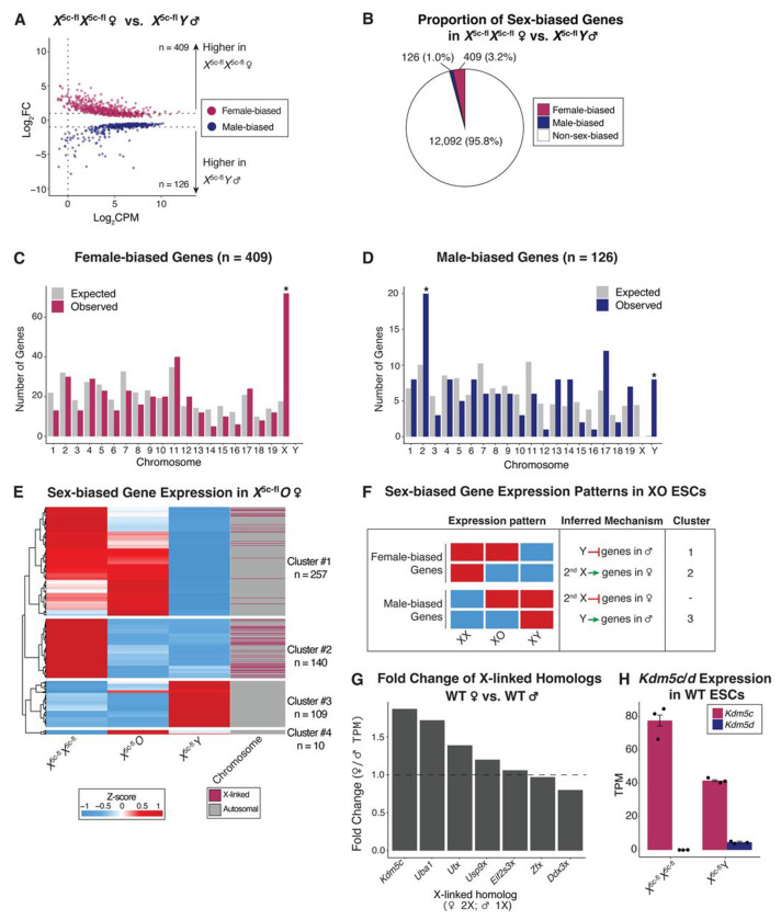
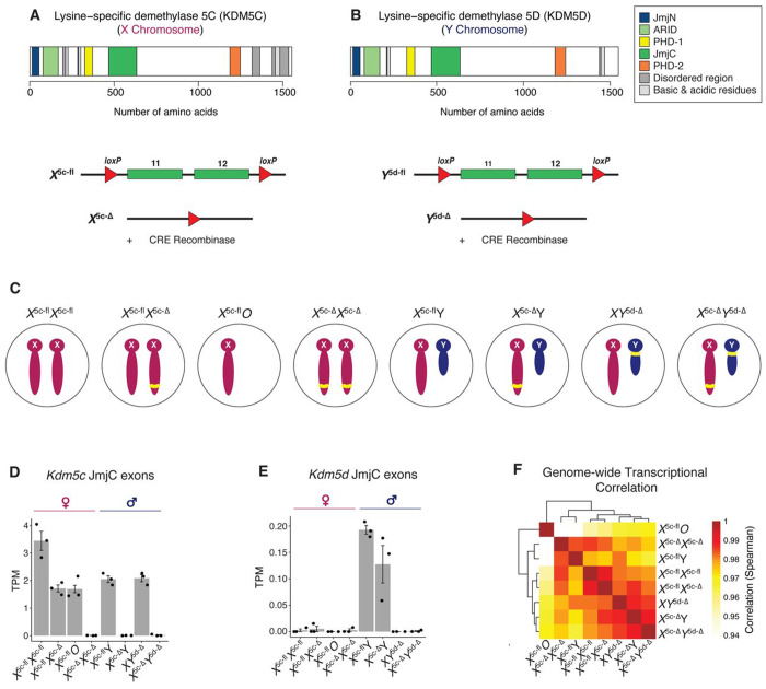
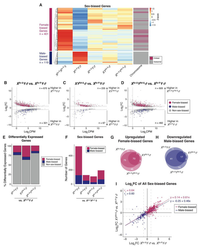
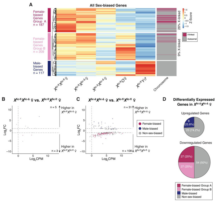
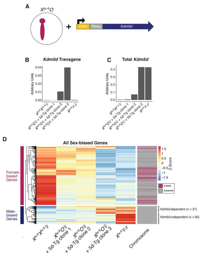
Conventionally, Y-linked Sry is thought to drive sex differences by triggering differential hormone production. Ancestral X-Y gene pairs, however, are hypothesized to drive hormone-independent sex differences. Here, we show that the X-Y gene pair Kdm5c-Kdm5d regulates sex-biased gene expression in pluripotent mouse embryonic stem cells (ESCs). Wild-type (WT) XX female ESCs exhibit >2-fold higher expression of 409 genes relative to WT XY male ESCs. Conversely, WT XY male ESCs exhibit >2-fold higher expression of 126 genes compared to WT XX female ESCs. Loss of Kdm5c in female ESCs downregulates female-biased genes. In contrast, loss of either Kdm5c or Kdm5d in male ESCs upregulates female-biased genes and downregulates male-biased genes, effectively neutralizing sex-biased gene expression. KDM5C promotes the expression of Kdm5d and several other Y-linked genes in male ESCs. Moreover, ectopic Kdm5d expression in female ESCs is sufficient to drive male-biased gene expression. These results establish Kdm5c-Kdm5d as critical regulators of sex-biased gene expression.

Keywords: Embryonic stem cells; Pluripotency; Sex chromosomes; Sex differences; X-Y gene pairs.


Conflict of interest statement

Declaration of interests S.I. is a member of the Scientific Advisory Board of KDM5C Advocacy, Research, Education & Support (KARES). The other authors declare no competing interests.

Figures

Figure 1:
Figure 1:. Sex-biased gene expression in WT X5c-flX5c-fl female WTand WT X5c-flY male ESCs.
(A) RNA-seq MA plot comparing WT X5c-flX5c-fl female vs. WT X5c-flY male ESCs (3 biological replicates each). ‘n’; number of differentially expressed genes (≥ 1 Log2 fold change (Log2FC), ≥ 1 count per million (CPM), false discovery rate (FDR) < 0.01). Maroon, female-biased genes. Dark blue, male-biased genes. Grey, non-sex-biased genes. Only genes with an FDR < 0.01 were plotted. (B) Proportion of expressed genes (CPM ≥ 1) in WT X5c-flX5c-fl female and WT X5c-flY male ESCs that exhibit sex-biased expression. (C and D) Distribution of female-biased (C) or male-biased (D) genes on each chromosome. Expected values were determined by Chi-square test. (*); p < 0.05 (binomial test, Benjamini-Hotchberg corrected) for difference between expected and observed number of sex-biased genes. (E) Heatmap of sex-biased gene expression (TPM) in XX, XO, and XY ESCs. Heatmap is clustered and scaled by row and colored by Z-score. Cluster cut-offs were determined by tree height (> 2). (F) Expression patterns of sex-biased genes in XX, XO, and XY ESCs and the inferred mechanisms by which the X and the Y chromosomes influence sex-biased gene expression. Cluster numbers correspond to (E). (G) Relative X-linked homolog expression (TPM) in WT X5c-flX5c-fl female vs. WT X5c-flY male ESCs. (H) Kdm5c and Kdm5d RNA expression in the WT X5c-flX5c-fl female vs. WT X5c-flY male ESCs (TPM; mean ± SEM).
Figure 2:
Figure 2:. Generation of Kdm5c and Kdm5d floxed and mutant ESCs.
(A and B) Conditional mutation strategy of Kdm5c (A; ref. 37) and Kdm5d (B; this study). In both cases, exons encoding the enzymatic JmjC domain were floxed (fl) and conditionally deleted (Δ) via germline Cre recombinase. (C) Schematic of all Kdm5c and Kdm5d WT and mutant male and female ESC lines used in this study. Yellow bar, Kdm5c mutation on the X chromosome or Kdm5d mutation on the Y chromosome. (D and E) Expression (TPM) of Kdm5c (D) or Kdm5d (E) JmjC targeted exons in the WT and mutant ESCs (mean ± SEM; 3 biological replicates each). (F) Spearman correlations of the transcriptomes (TPM) of all WT and mutant ESCs. Heatmap is scaled by row and colored by Z-score.
Figure 3:
Figure 3:. Neutralization of sex-biased gene expression in Kdm5c- and Kdm5d-mutant male ESCs.
(A) Heatmap of sex-biased gene expression (TPM) in WT X5c-flX5c-fl female and WT X5c-flY male ESCs vs. X5c-ΔY, XY5d-Δ, and X5c-Δ Y5d-Δ mutant male ESCs. Heatmap is clustered by row, scaled by row, and colored by Z-score. Cluster cut-offs were determined by tree height (≥ 3.5). (B-D) RNA-seq MA plots of pairwise comparisons of X5c-ΔY (B), XY5d-Δ (C), and X5c-Δ Y5d-Δ (D)mutant male ESCs vs. WT X5c-flY male ESCs. Maroon, female-biased genes. Dark blue, male-biased genes. Grey, non-sex-biased genes. Only genes with an FDR < 0.01 were plotted. (E)The proportions of differentially expressed genes in X5c-ΔY, XY5d-Δ, and X5c-Δ Y5d-Δ mutant male ESCs that exhibit sex-biased expression. (F) Number of sex-biased genes determined by pairwise comparisons between WT X5c-flX5c-fl female ESCs vs. X5c-ΔY, XY5d-Δ, and X5c-Δ Y5d-Δ mutant male ESCs. (G-H) Overlap of differentially expressed female-biased genes (G) and male-biased genes (H) in X5c-ΔY, XY5d-Δ, and X5c-Δ Y5d-Δ mutant male ESCs. (I) Correlation of sex-biased gene expression Log2FCs in X5c-ΔY and XY5d-Δ mutant male ESCs. Maroon, female-biased genes. Dark blue, male-biased genes. Linear models were determined by y ~ x. rs, Spearman correlation. Three biological replicates were used per genotype and all ‘n’ numbers indicate genes that were filtered for ≥ 1 Log2FC, ≥ 1 CPM, and FDR < 0.01.
Figure 4:
Figure 4:. Reduced sex-biased gene expression in Kdm5c-mutant female ESCs.
(A) Heatmap of sex-biased gene expression (TPM) in WT X5c-flX5c-fl female and WT X5c-flY male vs. Kdm5c-mutant female ESCs. Heatmap is clustered by row, scaled by row, and colored by Z-score. Cluster cut-offs were determined by tree height (≥ 3). (B-C) RNA-seq MA plots of pairwise comparisons of X5c-flX5c-Δ heterozygous mutant female (B) and X5c-ΔX5c-Δ homozygous mutant female ESCs (C) vs. WT X5c-flX5c-fl female ESCs. Maroon; female-biased genes. Dark blue; male-biased genes. Grey; non-sex-biased genes. Only genes with an FDR < 0.01 were plotted. (D) Proportion of upregulated or downregulated genes in X5c-ΔX5c-Δ homozygous mutant female ESCs that are sex-biased in expression. Three biological replicates were used per genotype and all ‘n’ numbers indicate genes that were filtered for ≥ 1 Log2FC, ≥ 1 CPM, and FDR < 0.01.
Figure 5:
Figure 5:. Kdm5d is sufficient to induce male-biased gene expression in X5c-flO ESCs.
(A) Schematic depicting experimental approach. A Kdm5d transgene tagged at the N-terminus with a Strep tag and constitutively driven by a CMV promoter was introduced into X5c-flO ESC clones by lentivirus. (B) The RNA expression levels of the Kdm5d transgene determined by RT-qPCR with a forward primer specific to the Strep tag and a reverse primer in the 5’ end of Kdm5d. Data are normalized to Tbp RNA. (C) The RNA expression levels of total Kdm5d by RT-qPCR with primers specific to Kdm5d exons 11 and 12. Data are normalized to Tbp RNA. (D) Heatmap of sex-biased gene expression (TPM) in WT X5c-flX5c-fl female and WT X5c-flY male ESCs vs. X5c-flO Kdm5d-transgenic clone ESCs. Heatmap is clustered by row, scaled by row, and colored by Z-score. Cluster cut-offs were determined by tree height (≥ 4).

Similar articles

References

    1. Larney C., Bailey T.L., and Koopman P. (2014). Switching on sex: transcriptional regulation of the testis-determining gene Sry. Development 141, 2195–2205. 10.1242/dev.107052. - DOI - PMC - PubMed
    1. Arnold A.P. (2009). The organizational-activational hypothesis as the foundation for a unified theory of sexual differentiation of all mammalian tissues. Horm Behav 55, 570–578. 10.1016/j.yhbeh.2009.03.011. - DOI - PMC - PubMed
    1. Blencowe M., Chen X., Zhao Y., Itoh Y., McQuillen C.N., Han Y., Shou B.L., McClusky R., Reue K., Arnold A.P., and Yang X. (2022). Relative contributions of sex hormones, sex chromosomes, and gonads to sex differences in tissue gene regulation. Genome Res 32, 807–824. 10.1101/gr.275965.121. - DOI - PMC - PubMed
    1. Wilson C.A., and Davies D.C. (2007). The control of sexual differentiation of the reproductive system and brain. Reproduction 133, 331–359. 10.1530/REP-06-0078. - DOI - PubMed
    1. Mamsen L.S., Ernst E.H., Borup R., Larsen A., Olesen R.H., Ernst E., Anderson R.A., Kristensen S.G., and Andersen C.Y. (2017). Temporal expression pattern of genes during the period of sex differentiation in human embryonic gonads. Sci Rep 7, 15961. 10.1038/s41598-017-15931-3. - DOI - PMC - PubMed

Publication types