[Preprint]. 2024 Oct 23:2024.10.23.619891.
doi: 10.1101/2024.10.23.619891.

Convergent olfactory circuits for courtship in Drosophila revealed by ds-Tango

Affiliations

Convergent olfactory circuits for courtship in Drosophila revealed by ds-Tango

John D Fisher et al. bioRxiv. .

Abstract

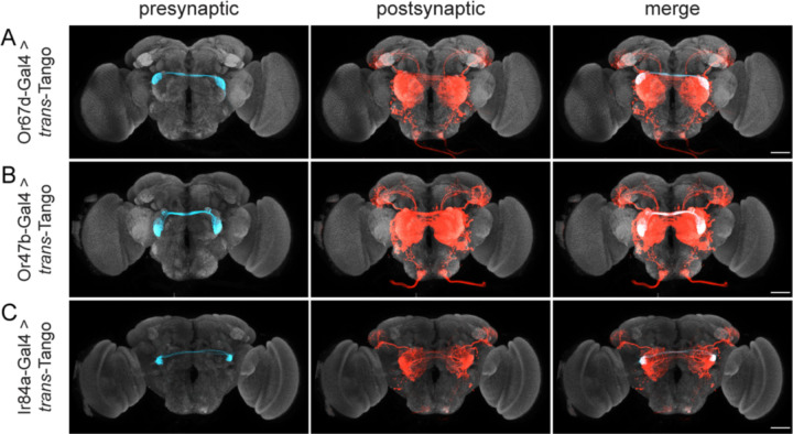
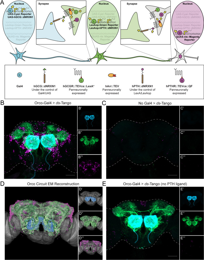
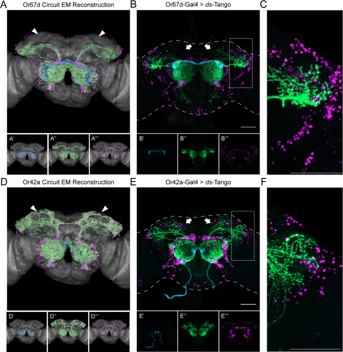
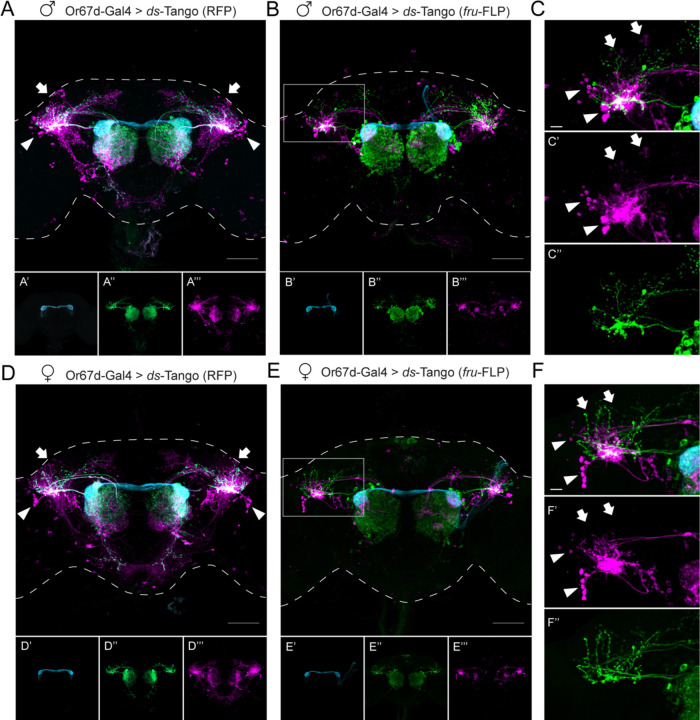
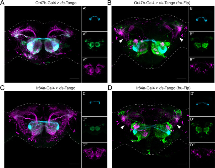
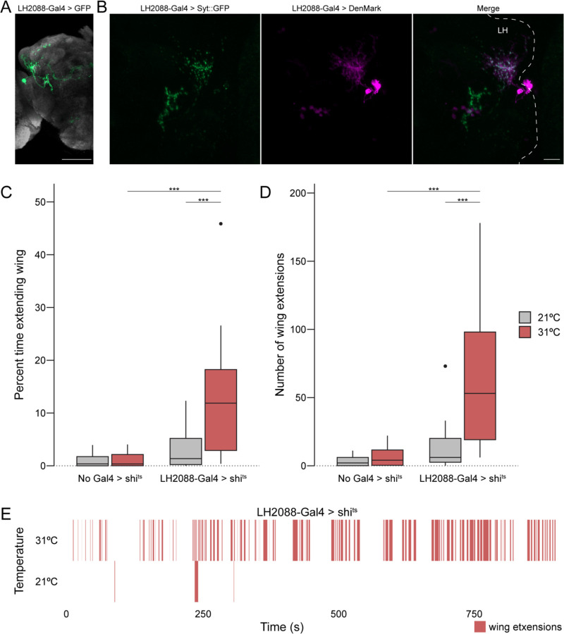
Animals exhibit sex-specific behaviors that are governed by sexually dimorphic circuits. One such behavior in male Drosophila melanogaster, courtship, is regulated by various sensory modalities, including olfaction. Here, we reveal how sexually dimorphic olfactory pathways in male flies converge at the third-order, onto lateral horn output neurons, to regulate courtship. To achieve this, we developed ds-Tango, a modified version of the monosynaptic tracing and manipulation tool trans-Tango. In ds-Tango, two distinct configurations of trans-Tango are positioned in series, thus providing selective genetic access not only to the monosynaptic partners of starter neurons but also to their disynaptic connections. Using ds-Tango, we identified a node of convergence for three sexually dimorphic olfactory pathways. Silencing this node results in deficits in sex recognition of potential partners. Our results identify lateral horn output neurons required for proper courtship behavior in male flies and establish ds-Tango as a tool for disynaptic circuit tracing.

Keywords: Drosophila; courtship; disynaptic tracing; ds-Tango; lateral horn; neural circuit.


Figures

Figure 1.
Figure 1.. Tracing second-order projections within the sexually dimorphic olfactory circuits
trans-Tango is used to trace the projections of the three FruM+ OSN populations: Or67d-OSNs (A), Or47b-OSNs (B), and Ir84a-OSNs (C). In the presynaptic channel (left column), only the OSNs are labeled (cyan). In the postsynaptic channel (center), both the LNs and PNs (red) are revealed. OPN axons target ventral LH in (A) and (B), and both ventral and dorsal LH in (C). Merge of both channels is shown in the rightmost column. Maximum intensity Z-stack projection of whole-mount brains are shown. Neuropil counterstain is shown in grey. Scale bars, 50μm.
Figure 2.
Figure 2.. Design and implementation of ds-Tango in the olfactory system
(A) Schematic and components of ds-Tango. In flies carrying a Gal4 driver, the presynaptic reporter (cyan) and the GCG ligand (hGCG::dNRXN1) are expressed in the starter neurons. The GCG ligand localizes to the presynaptic sites of the starter neurons and activates the GCGR Tango fusion (hGCGR::TEVcs::LexA*) across the synapse on the monosynaptic partners. Upon activation of the GCGR, the hArr::TEV fusion protein is recruited to it, TEV cleaves its recognition site (TEVcs) releasing LexA*. LexA* then translocates to the nucleus and initiates the expression of the PTH ligand (hPTH::dNRXN1) and of the monosynaptic reporter (green). The PTH ligand localizes to the presynaptic sites of the monosynaptic partners and activates the PTHR Tango fusion (hPTHR::TEVcs::QF) across the synapse on the disynaptic connections. Upon activation of the PTHR, the hArr::TEV fusion protein is recruited to it, TEV cleaves its recognition site (TEVcs), releasing QF. QF then translocates to the nucleus and initiates the expression of the nuclear disynaptic reporter (magenta). The various steps in the process are indicated by yellow arrows. (B) Driving ds-Tango in the peripheral olfactory system using Orco-Gal4 labels OSNs (cyan, shown in B’), their monosynaptic partners LNs and OPNs (green, shown in B’’), and the nuclei of their disynaptic connections (magenta, shown in B’’’). (C) A brain of a control fly bearing the ds-Tango components, but no Gal4 driver exhibits no background neurons in the presynaptic channel (cyan, shown in C’), virtually no background neurons in the monosynaptic channel (green, shown in C’’), and the nuclei of a few background neurons in the disynaptic channel (magenta, shown in C’’’). (D) EM reconstruction of the Orco circuit projected on a template brain (gray) reveals OSNs (cyan, shown in D’), LNs and OPNs (green, shown in D’’), and the nuclei of third-order olfactory neurons (magenta, shown in D’’’). (E) A brain of a control fly bearing Orco-Gal4 and a version of ds-Tango lacking the PTH ligand exhibits labeling in OSNs (cyan, shown in E’), LNs and OPNs (green, shown in E’’), but no staining of disynaptic partners except for the nuclei of a few background neurons (magenta, shown in E’’’). Maximum intensity Z-stack projection of whole-mount brains are shown in B, D, and E. Dashed lines in B, D, and E depict the approximate outline of the fly brains. Scale bars = 50 μm.
Figure 3.
Figure 3.. Specificity of ds-Tango
(A) Or67d Circuit EM reconstruction projected on a template brain (gray) reveals the OSNs (cyan, shown in A’), LNs and OPNs (green, shown in A’’), and the nuclei of third-order neurons (magenta, shown in A’’’). (B) Driving ds-Tango using Or67d-Gal4 labels Or67d-OSNs (cyan, shown in B’), their monosynaptic partner LNs and OPNs (green, shown in B’’), and the nuclei of their disynaptic connections (magenta, shown in B’’’). (C) Higher magnification image of the gray inset in (B), depicting the lateral horn. (D) Or42a Circuit EM reconstruction projected on a template brain (gray) reveals the OSNs (cyan, shown in D’), LNs and OPNs (green, shown in D’’), and the nuclei of third-order neurons (magenta, shown in D’’’). (E) Driving ds-Tango using Or42a-Gal4 labels Or42a-OSNs (cyan, shown in E’), their monosynaptic partners LNs and OPNs (green, shown in E’’), and the nuclei of their disynaptic connections (magenta, shown in E’’’). (F) Higher magnification image of the gray inset in (E), depicting the lateral horn. Arrowheads in A and D highlight the absence and presence of third-order Kenyon cells in the MB, respectively. Arrows in B and E highlight the absence and presence of third-order Kenyon cells in the MB, respectively. Maximum intensity Z-stack projection of whole-mount brains are shown in B and E. Dashed lines in B and E depict the approximate outline of the fly brains. Scale bars = 50 μm.
Figure 4.
Figure 4.. ds-Tango reveals sexual dimorphism in the lateral horn in the Or67d circuit
(A) Driving ds-Tango with Or67d-Gal4 in males labels Or67d-OSNs (cyan, shown in A’), their monosynaptic partner LNs and OPNs (green, shown in A’’), and their disynaptic connections (magenta, shown in A’’’). Arrows indicate the dorsal cluster neurons; arrowheads indicate the lateral cluster neurons. (B) Driving ds-Tango with Or67d-Gal4 and genetically restricting disynaptic reporter expression to fru-FLP+ neurons in males labels Or67d-OSNs (cyan, shown in B’), their monosynaptic partner LNs and OPNs (green, shown in B’’), and their fru-FLP+ disynaptic connections (magenta, shown in B’’’). (C) A higher magnification image of the gray inset in (B) highlighting the left LH reveals PNs targeting the ventral region of the LH (green, shown in C’’), overlapping with the neurites of fru-FLP+ disynaptic connections (magenta, shown in C’). Note the presence of lateral cluster neurons (arrowheads) and the dorsal cluster neurons (arrows) in the male brain. (D) Driving ds-Tango with Or67d-Gal4 in females labels Or67d-OSNs (cyan, shown in D’), their monosynaptic partner LNs and OPNs (green, shown in D’’), and their disynaptic connections (magenta, shown in D’’’). Arrowheads indicate the lateral cluster neurons; arrows indicate the location of dorsal cluster neurons. Note the presence of lateral cluster neurons in both the male (arrowheads in A) and female (arrowheads in D) brains and the prominent dorsal cluster neurons in the male (arrows in A) brain that are less prominent or absent in the female (arrows in D) brain. (E) Driving ds-Tango with Or67d-Gal4 and genetically restricting disynaptic reporter expression to fru-FLP+ neurons in females labels Or67d-OSNs (cyan, shown in E’), their monosynaptic partners LNs and OPNs (green, shown in E’’), and their fru-FLP+ disynaptic connections (magenta, shown in E’’’). (F) A higher magnification image of the gray inset in (E) showing the left LH reveals PNs targeting the ventral region of the LH (green, shown in F’’), overlapping with the neurites of fru-FLP+ disynaptic connections (magenta, shown in F’). Note the presence of lateral cluster neurons (arrowheads) and the absence of dorsal cluster neurons (arrows) in the female brain. Maximum intensity Z-stack projection of whole-mount brains are shown in A, B, D, and E. Dashed lines in A, B, D, and E depict the approximate outline of the fly brains. Scale bars, 50 μm.
Figure 5.
Figure 5.. A node of convergence for courtship-regulating olfactory circuits
(A) Driving ds-Tango with Or47b-Gal4 in males labels Or47b-OSNs (cyan, shown in A’), their monosynaptic partner LNs and OPNs (green, shown in A’’), and their disynaptic connections (magenta, shown in A’’’). (B) Driving ds-Tango with Or47b-Gal4 and genetically restricting disynaptic reporter expression to fru-FLP+ neurons in males labels Or47b-OSNs (cyan, shown in B’), their monosynaptic partner LNs and OPNs (green, shown in B’’), and their fru-FLP+ disynaptic connections (magenta, shown in B’’’). Note the presence of ventral projections from the LH to the AVLP in the disynaptic connections characteristic of AV2b1/b2 neurons (arrowheads). (C) Driving ds-Tango with Ir84a-Gal4 in males labels Ir84a-OSNs (cyan, shown in C’), their monosynaptic partners LNs and OPNs (green, shown in C’’), and their disynaptic connections (magenta, shown in C’’’). (D) Driving ds-Tango with Ir84a-Gal4 and genetically restricting disynaptic reporter expression to fru-FLP+ neurons in males labels Ir84a-OSNs (cyan, shown in D’), their monosynaptic partners LNs and OPNs (green, shown in D’’), and their fru-FLP+ disynaptic connections (magenta, shown in D’’’). Note the presence of ventral projections from the LH to the AVLP in the disynaptic connections characteristic of AV2b1/b2 neurons (arrowheads). Maximum intensity Z-stack projection of whole-mount brains are shown. Dashed lines depict the approximate outline of the fly brains. Scale bars, 50 μm.
Figure 6.
Figure 6.. Anatomical and functional analysis of the Maiandros neurons
(A) Expression pattern of the LH2088-Gal4 driver line. LH2088-Gal4 drives UAS-GFP (green). The Maiandros neurons are clearly labeled along with minimal background in the MB. Neuropil counterstain is shown in grey. (B) Locations of the Maiandros neuron axons and dendrites. LH2088-Gal4 drives synaptic vesicle marker Syt::GFP (green) and dendritic marker DenMark (magenta). Axons and dendrites are intermixed in the LH while ventral neurites in the AVLP are primarily axons. Dashed lines indicate the outline of the central brain. LH indicates lateral horn. (C) Quantification of unilateral wing extension as indicator of attempted courtship when each experimental male fly is paired with a single naïve WT male. Boxplot depicting the percent of total trial time (15 min) that experimental male flies extended one wing during thermogenetic silencing using shits. Silencing of the Maiandros neurons in male flies tested at the restrictive temperature of 31°C results in increased time spent with one wing extended relative to genetic controls and to experimental flies tested at the permissive temperature of 21°C. (D) Quantification of unilateral wing extension as indicator of attempted courtship when each experimental male fly is paired with a single naïve WT male. Boxplot depicting the total number of unilateral wing extensions exhibited by experimental male flies during thermogenetic silencing using shits. Silencing of the Maiandros neurons in male flies tested at the restrictive temperature of 31°C results in increases in total number of wing extensions relative to genetic controls and to experimental flies tested at the permissive temperature of 21°C. (E) Rasterplot depicting bouts of unilateral wing extensions over time for representative trials of male flies bearing the LH2088-Gal4 and UAS-shits alleles in the thermogenetic silencing assay at the permissive temperature of 21°C and the restrictive temperature of 31°C. Unilateral wing extensions are sparse at 21°C but abundant throughout the 31°C trial. Maximum intensity Z-stack projection of whole-mount brains are shown in (A) and (B). Scale bars, 50μm in (A) and 10μm in (B).

References

    1. Wong R.Y., and Hopkins C.D. (2007). Electrical and behavioral courtship displays in the mormyrid fish Brienomyrus brachyistius. J Exp Biol 210, 2244–2252. 10.1242/jeb.003509. - DOI - PubMed
    1. Bohn K.M., Schmidt-French B., Ma S.T., and Pollak G.D. (2008). Syllable acoustics, temporal patterns, and call composition vary with behavioral context in Mexican free-tailed bats. J Acoust Soc Am 124, 1838–1848. 10.1121/1.2953314. - DOI - PMC - PubMed
    1. Miles M.C., and Fuxjager M.J. (2018). Animal choreography of song and dance: a case study in the Montezuma oropendola,. Anim Behav 140, 99–107. 10.1016/j.anbehav.2018.04.006. - DOI
    1. Girard M.B., Elias D.O., and Kasumovic M.M. (2015). Female preference for multi-modal courtship: multiple signals are important for male mating success in peacock spiders. Proc Biol Sci 282, 20152222. 10.1098/rspb.2015.2222. - DOI - PMC - PubMed
    1. Greenspan R.J., and Ferveur J.F. (2000). Courtship in Drosophila. Annu Rev Genet 34, 205–232. 10.1146/annurev.genet.34.1.205. - DOI - PubMed

Publication types

LinkOut - more resources