[Preprint]. 2024 Oct 22:2024.10.22.619703.
doi: 10.1101/2024.10.22.619703.

Arc mediates intercellular tau transmission via extracellular vesicles

Affiliations

Arc mediates intercellular tau transmission via extracellular vesicles

Mitali Tyagi et al. bioRxiv. .

Abstract

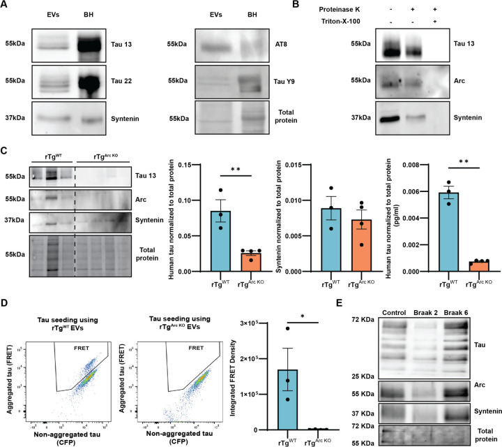
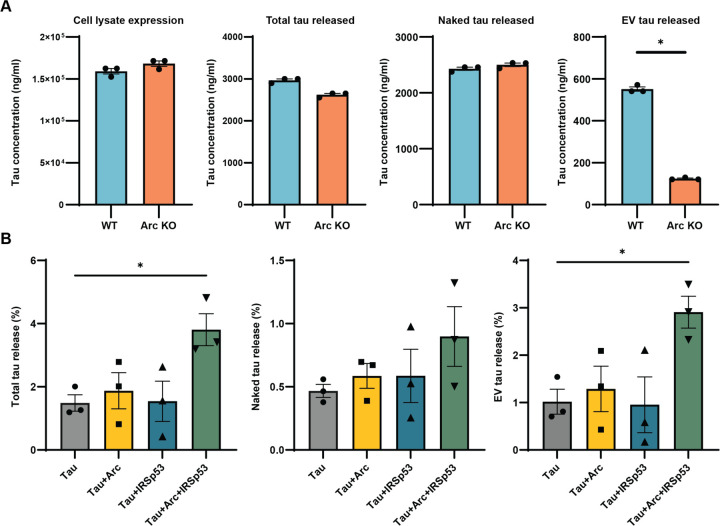
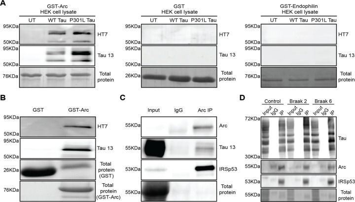
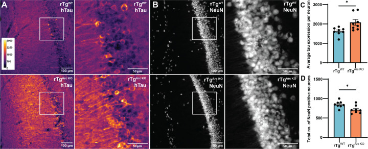
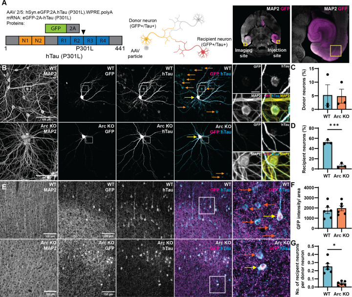
Intracellular neurofibrillary tangles that consist of misfolded tau protein1 cause neurodegeneration in Alzheimer's disease (AD) and frontotemporal dementia (FTD). Tau pathology spreads cell-to-cell2 but the exact mechanisms of tau release and intercellular transmission remain poorly defined. Tau is released from neurons as free protein or in extracellular vesicles (EVs)3-5 but the role of these different release mechanisms in intercellular tau transmission is unclear. Here, we show that the neuronal gene Arc is critical for packaging tau into EVs. Brain EVs purified from human tau (hTau) transgenic rTg4510 mice (rTgWT) contain high levels of hTau that are capable of seeding tau pathology. In contrast, EVs purified from rTgWT crossed with Arc knock-out mice (rTgArc KO) have significantly less hTau and cannot seed tau aggregation. Arc facilitates the release of hTau in EVs produced via the I-BAR protein IRSp53, but not free tau. Arc protein directly binds hTau to form a fuzzy complex that we identified in both mouse and human brain tissue. We find that pathological intracellular hTau accumulates in neurons in rTgArc KO mice, which correlates with accelerated neuron loss in the hippocampus. Finally, we find that intercellular tau transmission is significantly abrogated in Arc KO mice. We conclude that Arc-dependent release of tau in EVs plays a significant role in intracellular tau elimination and intercellular tau transmission.


Figures

Figure 1.
Figure 1.. Arc is critical for the release of seed-competent hTau in EVs.
A. hTau is released in brain EVs isolated from rTgWT mice. EVs were isolated from brains of 4-month-old rTgWT mice (n=3M). EVs and brain homogenates (BH) from 4-month-old rTgWT mice were immunoblotted for hTau (Tau 13, Tau 22, AT8, and Tau Y9 antibodies) and Syntenin. Brain derived EVs contain hTau, including putative oligomeric forms as detected by Tau 13 and Tau 22 antibodies. B. hTau and Arc in EVs is protected from Proteinase K degradation. EVs were isolated from the brains of 4-month-old rTgWT mice (n=3F). Fractions 1–4 obtained after SEC were pooled and incubated with Proteinase K (7ug/ml) with or without detergent (1% triton-X-100) for 10 mins. A representative western blot shows Arc, hTau, and Syntenin are protected from Proteinase K degradation when no detergent is present. C. Arc facilitates the release of hTau in EVs isolated from rTg mice. EVs were isolated from the brains of 4-month rTgWT mice (n=6, 2M, 4F with each EV prep consisting of two mice) and rTgArc KO (n=8, 4M, 4F with each EV prep consisting of two mice). Fractions 1–4 were pooled and blotted for hTau (Tau 13), Arc, and Syntenin. (Dotted line indicates spliced blot to crop out irrelevant lanes). Levels of hTau were significantly reduced in the absence of Arc, while there was no difference in general EV production, as indicated by similar Syntenin levels. hTau levels in EVs isolated from rTgArc KO mice, as assessed by total hTau ELISA, are significantly reduced. D. EV-hTau is capable of tau seeding. The same EV samples used for hTau analysis were used in a FRET tau seeding assay. Equal amounts of rTgWT and rTgArcKO EVs were transfected in HEK biosensor cells and FRET positive cells were counted using flow cytometry. FRET positive HEK biosensor cells exhibiting hTau aggregation are observed in the FRET gate in the representative flow cytometry plots. There are fewer FRET positive cells that were transfected with rTgArc KO EVs as compared to FRET positive cells transfected with rTgWT EVs. The integrated FRET intensity (calculated by multiplying the percentage of FRET positive cells and median fluorescence intensity of FRET positive cells) of cells transfected with rTgArc KO EVs is significantly lower than cells transfected with rTgWT EVs, indicating a lack of tau seeding. E. Arc and hTau are present in EVs isolated from post-mortem human brain tissue. EVs were isolated from a control, Braak stage 2, and Braak stage 6 post-mortem human brain tissue (Brodmann area 8/9). Isolated EVs were immunoblotted for Arc, hTau (Tau 13 antibody), and Syntenin. Arc and hTau are expressed in human brain-derived EVs. (Statistical analysis: Unpaired t-test, *p<0.05, **p<0.01).
Figure 2.
Figure 2.. hTau is preferentially released in Arc-IRSp53 EVs.
A. WT and Arc KO primary cortical neurons were transduced with hSyn-eGFP-2A-hTau*P301L at DIV 7 and media was collected on DIV 18. hTau levels in the media were quantified with or without detergent (1% triton-X-100) using a hTau ELISA to quantify total hTau and naked hTau, respectively. A significant decrease in EV-hTau levels was observed in Arc KO neurons, while naked and total hTau release were similar to WT neurons. (Statistical analysis: Unpaired t-test, *p<0.05 – data points are technical replicates). B. hTau is released in Arc-IRSp53 EVs. Neuro2A cells were transfected with P301L hTau-Hibit alone or with Arc and/or IRSp53 for 24 hours. Conditioned media was collected 24 hours post full media change. Luminescence was measured with or without detergent to quantify total hTau or free hTau in the media. Free hTau release was not affected by Arc and/or IRSp53 expression. However, EV-hTau release was significantly enhanced when Arc and IRSp53 were co-expressed. (Statistical analysis: One-way ANOVA, *p<0.05 – data points are technical replicates).
Figure 3.
Figure 3.. Arc directly binds to hTau and forms a complex with IRSp53 in mouse and human cortex.
A. hTau binds Arc. GST-Arc, GST, or GST-Endophilin were immobilized on beads and incubated with HEK cell lysate from untransfected (UT), WT hTau (2N4R) transfected, or mutant (P301L) hTau (2N4R) transfected cells. hTau was not present in the eluted fractions collected from GST or GST-Endophilin but was detected in fractions collected from GST-Arc. B. Arc directly binds hTau. GST or GST-Arc was immobilized on beads and incubated with purified recombinant hTau (P301L). hTau was present in the eluted fraction collected from GST-Arc but not GST. C. hTau co-immunoprecipitates with Arc and IRSp53 in mouse cortex. Cortical tissue was collected from 4-month-old rTgWT mice that experienced 6 hours of enrichment to increase Arc expression. Brain homogenates were incubated with IgG (rabbit polyclonal, EMD Millipore) or Arc antibody (rabbit polyclonal, Synaptic systems). Input and IP samples were blotted for Arc (mouse monoclonal, Santa Cruz), hTau (mouse, Tau13 antibody), and IRSp53 (mouse, Abcepta). hTau was detected in Arc-IPs but not in IgG-IPs. D. Arc and IRSp53 co-immunoprecipitates with hTau in human post-mortem prefrontal cortical tissue. Human post-mortem brain tissue from control, Braak stage 2, and Braak stage 6 AD patients were incubated with IgG (rabbit mouse antibody, Cell signaling) or D5D8N hTau antibody (rabbit mouse antibody, Cell signaling). Input and IP samples were blotted for hTau (mouse monoclonal, Tau 13 antibody, Biolegend), Arc (purified E5 alpaca recombinant nanobody), and IRSp53 (mouse monoclonal, Abcepta). Arc and IRSp53 were specifically detected in hTau-IPs.
Figure 4.
Figure 4.. Intracellular hTau accumulates in neurons and there are less neurons in the cell-body layer of dorsal CA1 hippocampus in rTgArc KO mice.
Brain slices were collected from 4-month-old rTgWT and rTgArc KO littermates (n=8, 3M, 5F). Brain slices of the CA1 region of the dorsal hippocampus were immunostained for hTau (Tau13 antibody) and NeuN. A. Representative images of hTau staining in rTgWT and rTgArc KO CA1. B. Representative images of NeuN staining in the same sections from A. C. Quantification of average hTau expression per neuron in the cell body layer. rTgArc KO mice have significantly higher intracellular hTau levels. D. Quantification of the total number of NeuN positive neurons in dorsal CA1. rTgArc KO mice have significantly lower NeuN positive cells in the CA1 cell body layer. (Statistical analysis: Unpaired t-test, *p<0.05).
Figure 6.
Figure 6.. Arc is critical for intercellular hTau transmission.
A. Schematic of intercellular hTau transmission assay. AAV 2/5:hSyn.eGFP.2A.hTau*P301L produces equal amounts of eGFP and hTau (P301L) via a single mRNA cleaved at a P2A sequence. Transduced “donor neurons” express both eGFP and hTau proteins. For in vivo studies. 6-month-old WT and Arc KO mice were injected with AAV2/5: hSyn-eGFP-2A-hTau*P301L virus unilaterally in medial entorhinal cortex. Ten weeks post-injection, the brains were collected, sectioned, and immunostained for eGFP and hTau (using Tau Y9 and Tau 13 antibodies). Imaging was performed in contralateral media entorhinal cortex. B. Representative images of hTau and eGFP staining in WT and Arc KO hippocampal primary neurons. Neurons were sparsely transduced at DIV 7. 14 days post-transduction, the neurons were fixed and immunostained for eGFP and hTau (using Tau Y9 and Tau 13 antibodies). Transduced donor neurons are indicated by yellow arrows and recipient neurons are indicated by orange arrows. C. Virus transduction is the same in WT and Arc KO primary neurons. The percentage of donor neurons is similar in WT and Arc KO neurons, indicating comparable viral transduction. D. Intercellular hTau transmission is reduced in Arc KO primary neurons. The percentage of recipient neurons is significantly reduced in Arc KO neurons (n=3 independent cultures). E. Representative images of hTau and eGFP staining in WT and Arc KO entorhinal cortex. Transduced donor neurons are indicated by yellow arrows and recipient neurons are indicated by orange arrows. F. Virus transduction is the same in WT and Arc KO mice entorhinal cortex. WT and Arc KO mice show no significant differences in eGFP intensity normalized to area, indicating comparable levels of virus transduction in WT and Arc KO mice (n=6, 3M, 3F). G. Intercellular hTau transmission is reduced in Arc KO mice in vivo. The number of recipient neurons per donor neuron is significantly reduced in Arc KO mice, indicating reduced intercellular hTau transmission (n=6, 3M, 3F). (Statistical analysis: Unpaired t-test, *p<0.05, ***p<0.001).

Similar articles

References

    1. Annaert W. & De Strooper B. A cell biological perspective on Alzheimer’s disease. Annual review of cell and developmental biology 18, 25–51 (2002). 10.1146/annurev.cellbio.18.020402.142302 - DOI - PubMed
    1. Frost B., Jacks R. L. & Diamond M. I. Propagation of tau misfolding from the outside to the inside of a cell. J Biol Chem 284, 12845–12852 (2009). 10.1074/jbc.M808759200 - DOI - PMC - PubMed
    1. Fowler S. L. et al. Tau filaments are tethered within brain extracellular vesicles in Alzheimer’s disease. bioRxiv, 2023.2004.2030.537820 (2023). 10.1101/2023.04.30.537820 - DOI - PMC - PubMed
    1. Ruan Z. et al. Alzheimer’s disease brain-derived extracellular vesicles spread tau pathology in interneurons. Brain 144, 288–309 (2021). 10.1093/brain/awaa376 - DOI - PMC - PubMed
    1. Bodart-Santos V. et al. Alzheimer’s disease brain-derived extracellular vesicles reveal altered synapse-related proteome and induce cognitive impairment in mice. Alzheimers Dement 19, 5418–5436 (2023). 10.1002/alz.13134 - DOI - PubMed

Publication types