[Preprint]. 2024 Oct 22:2024.10.21.619528.
doi: 10.1101/2024.10.21.619528.

Opto2P-FCM: A MEMS based miniature two-photon microscope with two-photon patterned optogenetic stimulation

Affiliations

Opto2P-FCM: A MEMS based miniature two-photon microscope with two-photon patterned optogenetic stimulation

Gregory L Futia et al. bioRxiv. .

Abstract

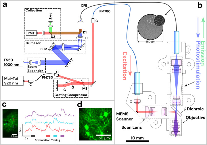
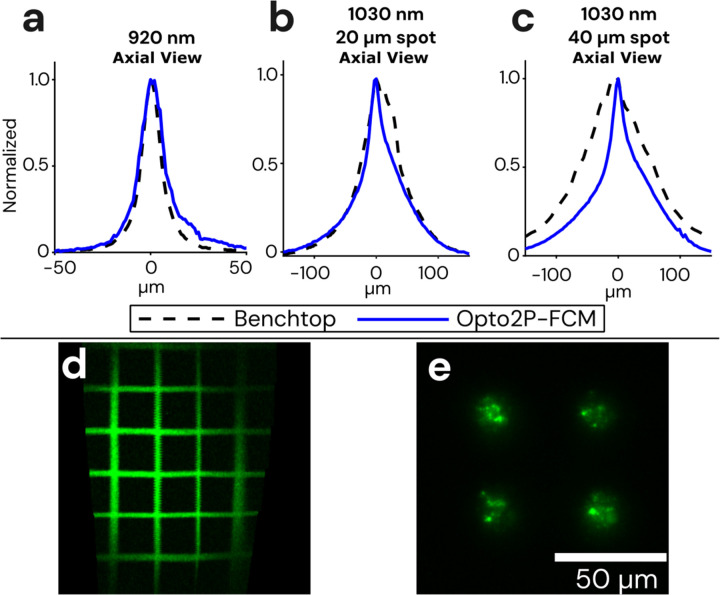
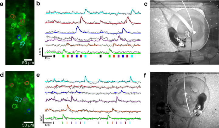
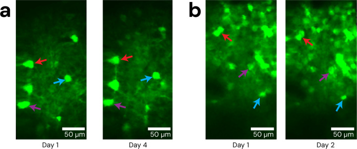
Multiphoton microscopy combined with optogenetic photostimulation is a powerful technique in neuroscience enabling precise control of cellular activity to determine the neural basis of behavior in a live animal. Two-photon patterned photostimulation has taken this further by allowing interrogation at the individual neuron level. However, it remains a challenge to implement imaging of neural activity with spatially patterned two-photon photostimulation in a freely moving animal. We developed a miniature microscope for high resolution two-photon fluorescence imaging with patterned two-photon optogenetic stimulation. The design incorporates a MEMS scanner for two-photon imaging and a second beam path for patterned two-photon excitation in a compact and lightweight design that can be head-attached to a freely moving animal. We demonstrate cell-specific optogenetics and high resolution MEMS based two-photon imaging in a freely moving mouse. The new capabilities of this miniature microscope design can enable cell-specific studies of behavior that can only be done in freely moving animals.


Conflict of interest statement

Competing financial interests K.K. is a co-founder and part-owner of 3i. The other authors declare no competing financial interests.

Figures

Figure 1:
Figure 1:. Diagram of Opto2P-FCM miniature two-photon microscope with optogenetic stimulation.
(a) Schematic of the lasers and detection system. The 920 nm pulsed laser is coupled into a polarization maintaining fiber (PM780) for spectral broadening and a transmission grating-pair compressor for dispersion compensation. The output is coupled to a second PM780 fiber to relay to the miniature microscope. A 1030 nm pulsed laser is beam-expanded and reflected off of a spatial light modulator (SLM) for holographic spatial patterning. The spatially patterned beam is focused onto a coherent imaging fiber bundle (CFB) to relay the patterned light to the miniature microscope. Fluorescence is collected back through the same CFB, separated by dichroics and detected on two photomultipliers (PMTs). (b) Schematic of miniature microscope with detailed layout of optical components and beam path. The 920 nm laser output from the PMF is collimated and reflected off of a MEMS scanner and passes through a scan lens consisting of a plano-convex and achromatic lens to generate a telecentric scan across the imaging plane. Patterned photostimulation light at 1030 nm exits the CFB, is collimated using an aspheric lens, combined with the 920 nm light using a dichroic mirror and focused through the same objective. The miniature objective is designed with a working distance of 1.5 mm to focus both 920 nm and 1030 nm light to the same imaging plane. (c) In vivo demonstration of head-attached two-photon imaging and selective optogenetic two-photon photostimulation in a freely moving mouse. Imaging was performed in the visual cortex with neurons co-expressing the calcium indicator jGCaMP7s and the opsin ChRmine. Two-photon photostimulation was performed on three selected regions of interest (ROI) in the imaging field (see outlines on the maximum intensity projection image). An image time sequence was acquired whereby all three ROIs were illuminated simultaneously (indicated by black stimulation timing bar) followed by each individual ROI sequentially (indicated by color matched bars). ΔF/F traces of jGCaMP7s show responses in the ROIs that occur synchronously with photostimulation. (d) The resolution of the Opto2P-FCM provides visualization of processes from somatosensory cortex neurons expressing jGCaMP7s.
Figure 2:
Figure 2:. Optical resolution and field of view measurements of the Opto2P-FCM.
(a) Axial resolution of the Opto2P-FCM was characterized by measuring the 2P fluorescence signal of a uniform flat fluorescent target scanned through the focus of the 920 nm laser (blue solid line, 14 μm FWHM) compared with a benchtop two-photon microscope with a 10x/0.4 NA objective (dashed line, 11 μm FWHM). Axial resolution of the 1030 nm patterned photostimulation was measured for a (b) 20 μm diameter circle using the Opto2P-FCM (blue solid line, 50 μm FWHM) compared to the benchtop microscope (dashed line, 54 μm FWHM) and for a (c) 40 μm diameter circle using the Opto2P-FCM (blue solid line, 54 μm FWHM) compared to the benchtop microscope (dashed line, 124 μm FWHM). (d) Opto2P-FCM image of a 50 μm grid slide showing a field of view of ~200 μm × 300 μm. (e) Image of photostimulation pattern (4 circles with 20 μm diameter) at the focus of the Opto2P-FCM recorded by 2P fluorescence from a thin rhodamine sample.
Figure 3:
Figure 3:. Demonstration of cell-specific 2P patterned photostimulation and simultaneous 2P imaging in a freely moving mouse.
The stimulation sequence consists of all ROIs together followed by stimulation of each ROI individually. Top panels were taken using a 0.5 m length CFB in visual cortex, with 5 ms/frame of stimulation for 2 seconds, bottom panels were taken using a 1 m length CFB in somatosensory cortex, with 10 ms/frame of stimulation for 700 ms. (a) and (d) show the fields and targeted/detected ROIs, with solid line circular ROIs indicating the targeted stimulation regions and dashed line ROIs indicating those detected by CaImAn. (b) and (e) show ΔF/F traces of jGCaMP7s activity during the recording timelapse color matched to ROIs in (a) and (d). Solid lines represent ΔF/F denoised traces from CaImAn and overlaid dashed lines are raw ΔF/F traces of the stimulation ROI. Color matched bars at the bottom represent stimulation times and durations, with black representing the stimulation of all ROIs. (c) and (f) show a still frame of the mouse during the recording. Behavioral video is shown in Video S1.
Figure 4:
Figure 4:. Repeatability of Opto2P-FCM imaging fields.
The Opto2P-FCM was head-attached to moving animals across multiple days, repeatably accessing the same cell field after being locked into the baseplate. (a) Example of repeated imaging of the same field across days in visual cortex and (b) somatosensory cortex. Colored arrows indicate several reference features.

Similar articles

References

    1. Shuman T. et al. Breakdown of spatial coding and interneuron synchronization in epileptic mice. Nat. Neurosci. 23, 229–238 (2020). - PMC - PubMed
    1. Guo C. et al. Miniscope-LFOV: A large-field-of-view, single-cell-resolution, miniature microscope for wired and wire-free imaging of neural dynamics in freely behaving animals. Sci. Adv. 9, eadg3918 (2023). - PMC - PubMed
    1. Hur S. W. et al. Correlated signatures of social behavior in cerebellum and anterior cingulate cortex. eLife 12, RP88439 (2024). - PMC - PubMed
    1. Cai D. J. et al. A shared neural ensemble links distinct contextual memories encoded close in time. Nature 534, 115–118 (2016). - PMC - PubMed
    1. Barbera G. et al. Spatially Compact Neural Clusters in the Dorsal Striatum Encode Locomotion Relevant Information. Neuron 92, 202–213 (2016). - PMC - PubMed

Publication types

LinkOut - more resources