[Preprint]. 2024 Oct 25:2024.10.22.617501.
doi: 10.1101/2024.10.22.617501.

Single-chain nanobody inhibition of Notch and avidity enhancement utilizing the β-pore forming toxin Aerolysin

Affiliations

Single-chain nanobody inhibition of Notch and avidity enhancement utilizing the β-pore forming toxin Aerolysin

Andrew C D Lemmex et al. bioRxiv. .

Update in

Abstract

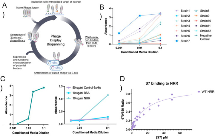
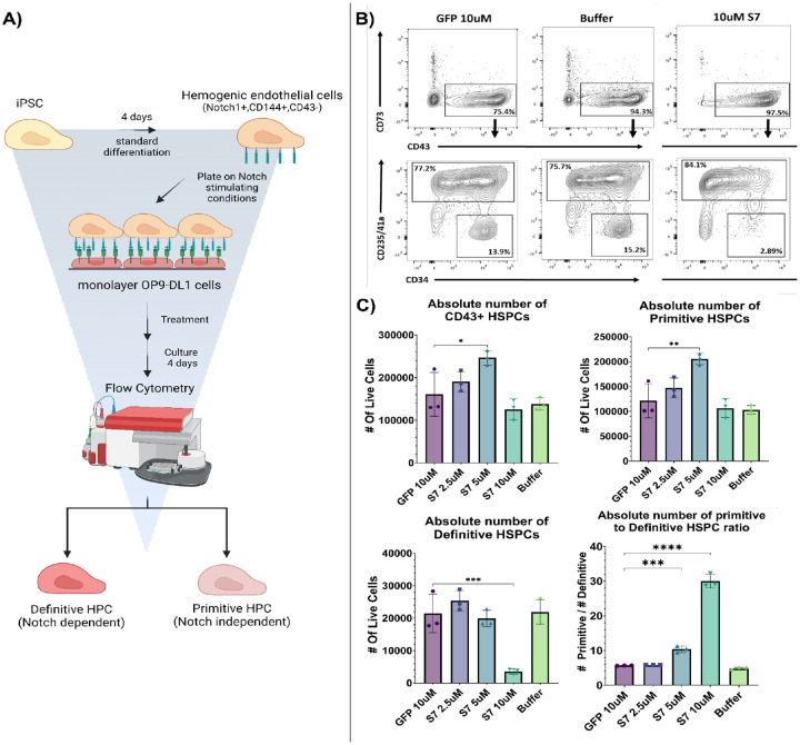
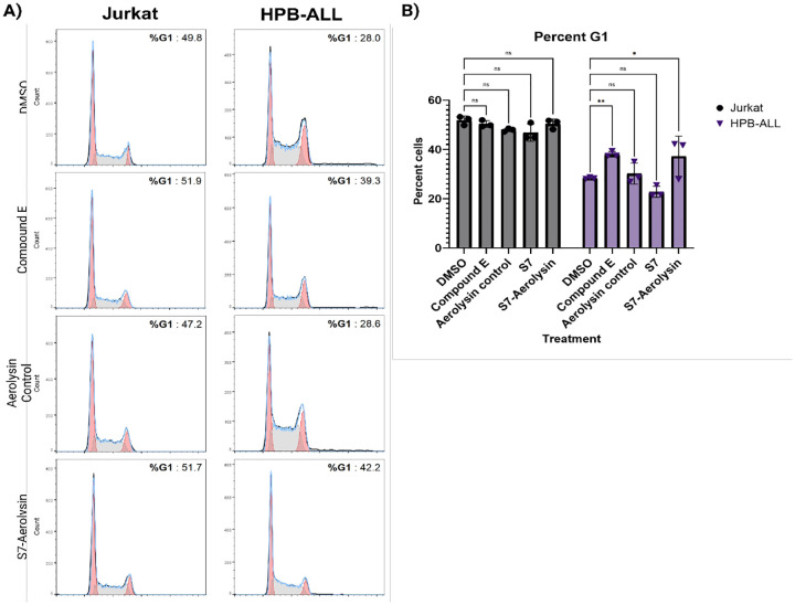
Notch plays critical roles in developmental processes and disease pathogenesis, which has led to numerous efforts to modulate its function with small molecules and antibodies. Here we present a nanobody inhibitor of Notch signaling, derived from a synthetic phage-display library targeting the notch Negative Regulatory Region (NRR). The nanobody inhibits Notch signaling in a luciferase reporter assay and in Notch-dependent hematopoietic progenitor cell differentiation assay, despite a modest 19uM affinity for Notch. We addressed the low affinity by fusion to a membrane-associating domain derived from the β-Pore forming toxin Aerolysin, resulting in a significantly improved IC50 for Notch inhibition. The nanobody-aerolysin fusion inhibits proliferation of T-ALL cell lines with similar efficacy to other Notch pathway inhibitors. Overall, this study reports the development of a Notch inhibitory antibody, and demonstrates a proof-of-concept for a generalizable strategy to increase the efficacy and potency of low-affinity antibody binders.


Conflict of interest statement

Conflict of interest statement The authors declare no conflicts of interest in this work.

Figures

Figure 1-
Figure 1-. Identification of a Notch1 NRR binding Nanobody.
(A) Anti-NRR ELISA of 12 candidates at 3 conditioned media dilutions. (B) Sanger sequencing of Strain 3, Strain 4, Strain 7, Strain 10, Strain 11, and Strain 12. Strain 10 contains an amber TAG codon which is read through in TG1 cells. (C) Testing of Strain 7 conditioned media on plates coated with an irrelevant protein M04–6xHis, BSA, or NRR. (D) Testing of Strain 7 conditioned media on plates coated with NRR or mClover3-NRR-mRuby3.
Figure 2-
Figure 2-. S7 inhibits Notch activation in cell-based assay.
(A) Schematic of signaling assay. Cells transfected with N1-Gal4 promoter are plated in plates precoated with Notch ligand. The Receptor binds the Notch ligand (1), leading to S2 and S3 proteolytic cleavages, releasing the intracellular Gal4 domain (2) and translocation to the nucleus where it drives transcription of firefly luciferase (3). Firefly luciferase signal is normalized against constitutive expression of Renilla luciferase. (B) Effect of serial dilution of S7 on Notch1-Gal4 signaling. Each point represents 3 technical replicates
Figure 3 –
Figure 3 –. S7 inhibits differentiation of hemogenic endothelial cells into definitive hematopoietic cells.
A. Assay scheme B. Flow cytometry contour plots showing relevant T-cell populations in the presence of buffer, GFP nanobody control, and S7 nanobody. C. Quantification of cell populations from panel B.
Figure 4 -
Figure 4 -. S7 fused to Aerolysin improves efficacy of S7 inhibitory effect.
(A) Cartoon representation of S7 fused to the N-terminus of Aerolysin and the localization of S7 to the surface of the cell via Aerolysin. (B) Cartoon representation of the assay in C, in which plates are coated with a titration of DLL4 ligand, cells with the Notch reporter are plated and then treated with a fixed concentration of S7 or S7-aerolysin to determine if the treatments shift the response to DLL4. (C) The response of a Notch reporter of activation in a cell-based signaling assay to treatment with either S7 or S7-Aerolysin at different concentrations. (D) The response of U2OS cells in the signaling assay plated on different concentrations of DLL4 ligand while treat with either 10uM S7 or 250nM S7-Aerolysin.
Figure 5–
Figure 5–. S7-Aerolsyin alters cell surface staining populations of Notch1
(A) Cartoon schematic of the experiment showing the doxycycline induction of the Notch1 receptor with a FLAG tag on the extracellular side, and a GFP fused to the C-terminus of the intracellular side. Cells are treated and then stained with an Anti-FLAG-APC antibody to stain Notch at the cell surface. Cells are then analyzed on flow, and the APC signal is normalized to the overall GFP signal to control for the total amount of Notch in the cell vs Notch just on the cell-surface. (B) Quantification of the Median RFU ratio between APC signal and GFP signal(n=3). Example dot plot of cells gated for APC and GFP signal, with the dotted red circle highlighting the shifted cell population (represented as colored dots) vs the induced population (represented as black dots). (D) Quantitation of the percent of total cell population that resides within the dotted red circle from panel C.
Figure 6 –
Figure 6 –. S7-Aerolysin inhibits the proliferation of T-ALL cell line sensitive to Notch-inhibition but does not alter proliferation of Notch-insensitive cell line.
(A) DNA staining and cell cycle analysis with Jurkate cells (Notch insensitive) vs HPB-ALL cells (Notch sensitive) with the indicated treatments. (B) Quantification of the percentage of cells in the G1-phase with the various treatments.

References

    1. Kovall R.A., Gebelein B., Sprinzak D., and Kopan R. (2017). The Canonical Notch Signaling Pathway: Structural and Biochemical Insights into Shape, Sugar, and Force. Dev. Cell 41, 228–241. - PMC - PubMed
    1. Bray S.J. (2006). Notch signalling: a simple pathway becomes complex. Nat. Rev. Mol. Cell Biol. 7, 678–689. - PubMed
    1. Shin B., Chang S.J., MacNabb B.W., and Rothenberg E.V. (2024). Transcriptional network dynamics in early T cell development. J. Exp. Med. 221. 10.1084/jem.20230893. - DOI - PMC - PubMed
    1. Zhou B., Lin W., Long Y., Yang Y., Zhang H., Wu K., and Chu Q. (2022). Notch signaling pathway: architecture, disease, and therapeutics. Signal Transduct. Target. Ther. 7, 95. - PMC - PubMed
    1. Gordon W.R., Zimmerman B., He L., Miles L.J., Huang J., Tiyanont K., McArthur D.G., Aster J.C., Perrimon N., Loparo J.J., et al. (2015). Mechanical Allostery: Evidence for a Force Requirement in the Proteolytic Activation of Notch. Dev. Cell 33, 729–736. - PMC - PubMed

Publication types

LinkOut - more resources