[Preprint]. 2024 Oct 22:2024.10.21.619433.
doi: 10.1101/2024.10.21.619433.

A rapid, facile, and economical method for the isolation of ribosomes and translational machinery for structural and functional studies

Affiliations

A rapid, facile, and economical method for the isolation of ribosomes and translational machinery for structural and functional studies

Jessey Erath et al. bioRxiv. .

Update in

Abstract

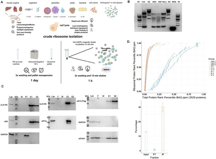
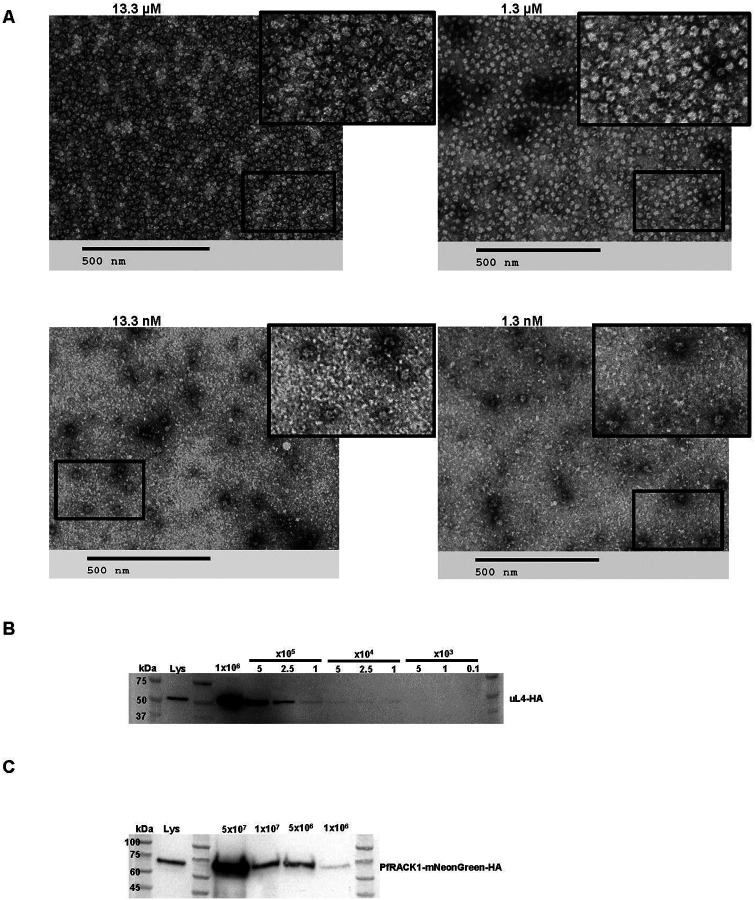
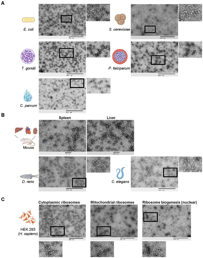
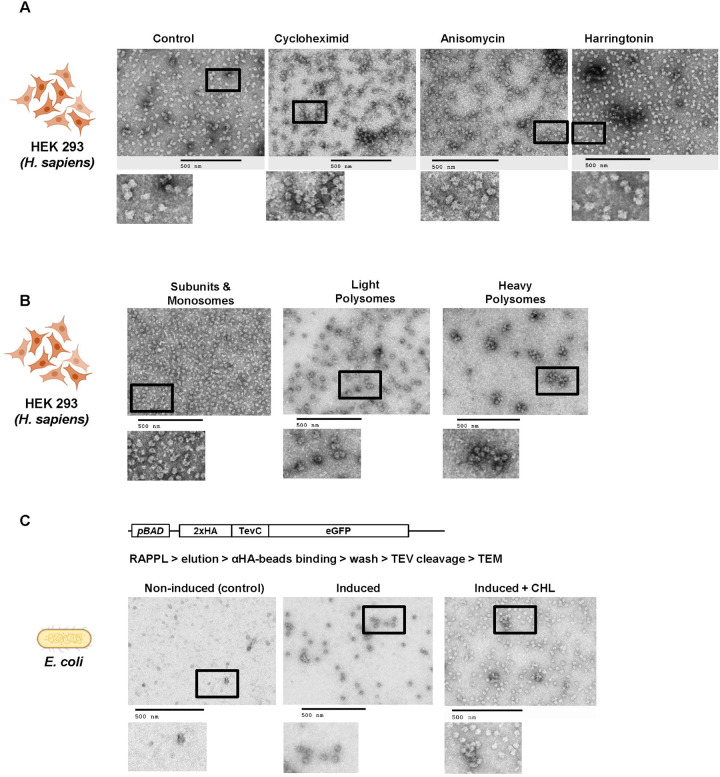
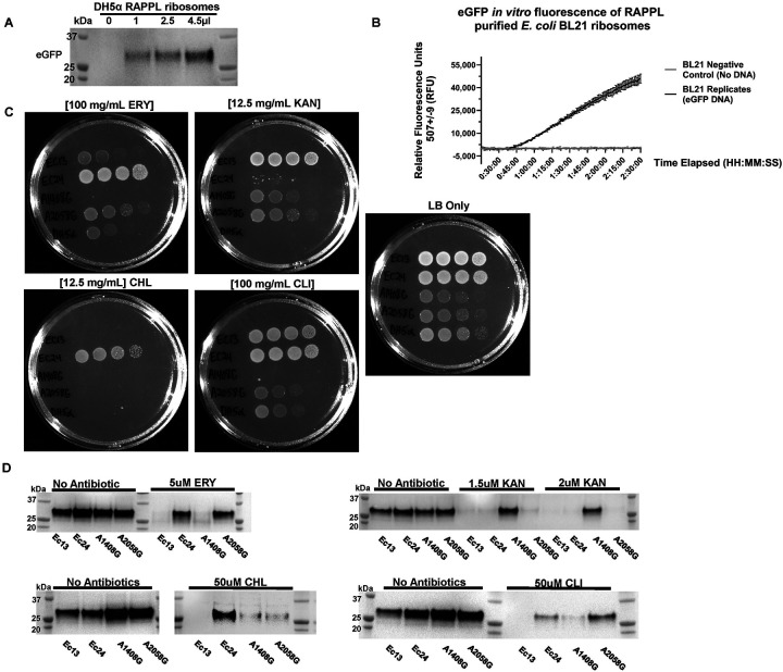
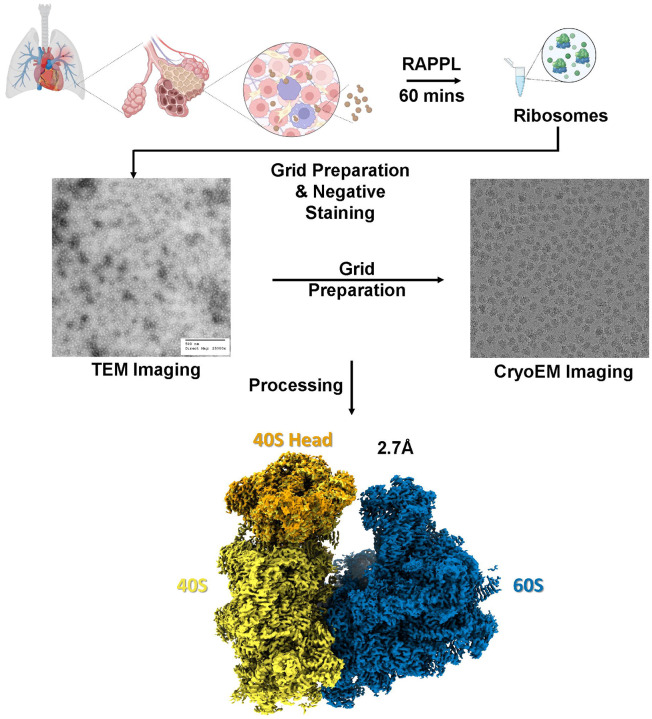
Ribosomes are macromolecular RNA-protein complexes that constitute the central machinery responsible for protein synthesis and quality control in the cell. Ribosomes also serve as a hub for multiple non-ribosomal proteins and RNAs that control protein synthesis. However, the purification of ribosomes and associated factors for functional and structural studies requires a large amount of starting biological material and a tedious workflow. Current methods are challenging as they combine ultracentrifugation, the use of sucrose cushions or gradients, expensive equipment, and multiple hours to days of work. Here, we present a rapid, facile, and cost-effective method to isolate ribosomes from in vivo or in vitro samples for functional and structural studies using single-step enrichment on magnetic beads - RAPPL (RNA Affinity Purification using Poly-Lysine). Using mass spectrometry and western blot analyses, we show that poly-lysine coated beads incubated with E. coli and HEK-293 cell lysates enrich specifically for ribosomes and ribosome-associated factors. We demonstrate the ability of RAPPL to isolate ribosomes and translation-associated factors from limited material quantities, as well as a wide variety of biological samples: cell lysates, cells, organs, and whole organisms. Using RAPPL, we characterized and visualized the different effects of various drugs and translation inhibitors on protein synthesis. Our method is compatible with traditional ribosome isolation. It can be used to purify specific complexes from fractions of sucrose gradients or in tandem affinity purifications for ribosome-associated factors. Ribosomes isolated using RAPPL are functionally active and can be used for rapid screening and in vitro characterization of ribosome antibiotic resistance. Lastly, we demonstrate the structural applications of RAPPL by purifying and solving the 2.7Å cryo-EM structure of ribosomes from the Cryptococcus neoformans, an encapsulated yeast causing cryptococcosis. Ribosomes and translational machinery purified with this method are suitable for subsequent functional or structural analyses and provide a solid foundation for researchers to carry out further applications - academic, clinical, or industrial - on ribosomes.


Figures

Fig. 1:
Fig. 1:. The RAPPL method.
A. Schematic describing the advantages of RAPPL over conventional methods. B. 2% agarose gel of RAPPLE purified RNA samples from of HEK-293 (HEK), human dermal fibroblasts (HDF) and HeLa human cell cultures. RNA isolated from commercial HeLa cell lysate (Con; Thermo Fisher HeLa IVT kit) and purified yeast tRNAs (tRNA, Ambion) were loaded as controls. NEB 100 bp and 1 Kb base pair markers (M1 and M2, respectively) are used to estimate size of isolated RNAs. C. Western blot analysis of HEK-293 lines uL4-HA and uS13-Flag tagged by CRISPR/Cas9 throughout the RAPPL purification process – lysate (Lys), flow-through (FT), wash (W) and elution (E1 and E2). RAPPL is selective for ribosome-associated factors, showing that the HA-tagged ribosomal protein uL4, Flag-tagged uS13, as well as the untagged uS9 are in the elution fractions. Translation factors are also enriched and purified by RAPPL as seen by the visualization of eIF3A and eIF4A1 proteins by specific antibodies in elution fractions. Presence of GAPDH, as a control for loading is detected in lysate and flow-through. Molecular markers indicate size of detected proteins. D. (top) Plot of the of each HEK-293 ribosomal protein’s rank percentile in relation to total protein rank percentile for each replicate of input, flow-through (FT), and bead-bound (IP) fractions. (bottom) Graph representing percentage of ribosomal proteins in total protein associated with input, flow-through (FT), and poly-lysine bead-bound (IP) fractions. Error bars represent standard deviation of triplicate averages.
Fig. 2:
Fig. 2:. RAPPL can enrich and purify ribosomes from limited biological material.
A. TEM visualization of RAPPL eluates from PureExpress® ribosomes from a 10-fold dilution scheme of 13.3 μM to 1.3 nM. The scale bar represents 500 nm. B. Western blot analysis using αHA antibody on RAPPL eluates of HEK-293 cells in which uL4 was HA-tagged by CRISPR/Cas9 whereby the starting cells were diluted to 1×10⁶, 5×10⁵, 2.5×10⁵, 1×10⁵, 5×10⁴, 2.5×10⁴, 1×10⁴, 5×10³, 1×10³, and 0.1×10³ cells prior to lysis. C. Western blot analysis using αHA antibody on RAPPL eluates of P. falciparum NF54 cells in which PfRACK was C-terminally tagged with mNeonGreen-HA whereby the starting cells were diluted to 5×10⁷, 1×10⁷, 5×10⁶, and 1×10⁶ cells. Molecular markers indicate size of detected proteins.
Fig. 3:
Fig. 3:. RAPPL is a versatile method multiple single celled organisms, tissues, and multicellular organisms.
A. TEM visualization of RAPPL eluates purified from several single celled organisms: E. coli, S. cerevisiae, T. gondii, P. falciparum, C. parvum. B. TEM visualization of RAPPL eluates purified from mouse tissue sections of spleen and liver as well as whole organisms D. rerio and C. elegans. C. TEM visualization of compartment-specific ribosomes generated from RAPPL eluates of cytoplasmic, mitochondrial, and nuclear (ribosome biogenesis) fractions. The scale bar represents 100 or 500 nm.
Fig. 4:
Fig. 4:. The elutions of RAPPL can be used in downstream applications.
A. HEK-293 lysates were treated with cycloheximide, anisomycine, and harringtonine with untreated lysate as a control. Ribosomes were purified using RAPPL in the presence of inhibitors and the eluates visualized by TEM. B. HEK-293 lysates were fractionated using polysome profiling. Fractions corresponding to ribosome subunit and monosomes, light polysomes, and heavy polysomes were pooled, respectively. These pools were diluted 1:5 to ensure that sucrose did not interfere with binding. The diluted, pooled samples were subject to RAPPL and the eluates visualized by TEM. C. Schematic of arabinose-inducible reporter expressing a 2xHA affinity tagged eGFP reporter separated by a TEV protease cleavage site (top). RAPPL was performed on bacterial lysates in the absence or presence of bacterial translation elongation inhibitor chloramphenicol (CHL) followed by αHA magnetic bead purification, again ±CHL, finally eluting with TEV protease. Eluates were visualized by TEM. Non-induced are shown as controls for lack of protein production and subsequent non-specific binding to αHA beads. The scale bar represents 500 nm.
Fig. 5:
Fig. 5:. RAPPL isolated ribosomes are translationally active and can be used for clinical applications.
A. Ribosomes were purified by RAPPL from E. coli DH5α cells grown to exponential phase, eluting in 30 μL of RAPPL elution buffer. Eluates were then used in the PURExpress® in vitro translation system instead of kit ribosomes. A PCR product encoding for eGFP harboring the T7 promoter and a polyA tail was used in the reaction (See Method for full details). Reactions were incubated for four hours. Ribosomes purified using RAPPL are active and able to translation mRNA. B. Activity of RAPPL purified E. coli BL21 ribosomes in the in vitro PURExpress® assays were observed using a kinetics protocol measuring eGFP fluorescence on an imaging plate reader. Relative fluorescence was determined with excitation settings set to wavelengths of 488 ± 9 and emission settings set to wavelengths of 507 ± 9. The standard deviation of technical triplicates eGFP fluorescence activity over a two hours and 30 minutes period with and without the DNA template encoding for eGFP are shown on the graph. C. Plate bacterial growth assays were performed using erythromycin (ERY), kanamycin (KAN), chloramphenicol (CHL), and clindamycin (CLI) to demonstrate strain resistance with LB only as controls for growth and the DH5α strain was used as a control strain. Concentration of used antibiotics is indicated. D. Synthesis of eGFP reporter by RAPPL isolated ribosomes in the absence and presence of indicated antibiotics (ERY, KAN, CHL, and CLI) targeting E. coli ribosomes. For each strain, 4.5 μL of 1.5 μg / μL of RAPPL isolated ribosomes was used for standard 25 μL PURExpress® in vitro Δ ribosome translation reaction (See Method for full details). Western blot analysis was performed on samples collected after 4 hours of incubation at 37°C and visualized using αGFP specific antibody. Molecular markers indicate size of eGFP protein.
Fig. 6:
Fig. 6:. Structural determination of RAPPL products can produce high-resolution CryoEM Maps.
C. neoformans cells (~108) in exponential phase were lysed and the clarified lysate used in RAPPL. The ribosomes were eluted in 30 μL of elution buffer. The Eluate was first screened using TEM. Subsequently, grids were prepared using 5 μL of eluate. Movies were captured on FEI Titan Krios G3 300kV Cryo-TEM with Falcon IV Direct Electron Detection camera. Data was processed using cryoSPARC resulting in an ~2.7 Å global resolution.

References

    1. Keller E. B., Zamecnik P. C., Loftfield R. B., The role of microsomes in the incorporation of amino acids into proteins. J Histochem Cytochem 2, 378–386 (1954). - PubMed
    1. Palade G. E., A small particulate component of the cytoplasm. J Biophys Biochem Cytol 1, 59–68 (1955). - PMC - PubMed
    1. Nirenberg M. W., Matthaei J. H., The dependence of cell-free protein synthesis in E. coli upon naturally occurring or synthetic polyribonucleotides. Proceedings of the National Academy of Sciences 47, 1588–1602 (1961). - PMC - PubMed
    1. Ban N., Nissen P., Hansen J., Moore P. B., Steitz T. A., The complete atomic structure of the large ribosomal subunit at 2.4 A resolution. Science 289, 905–920 (2000). - PubMed
    1. Moore P. B., Steitz T. A., The structural basis of large ribosomal subunit function. Annu Rev Biochem 72, 813–850 (2003). - PubMed

Publication types

LinkOut - more resources