[Preprint]. 2024 Oct 24:2024.10.22.619649.
doi: 10.1101/2024.10.22.619649.

Bioorthogonal cyclopropenones for investigating RNA structure

Affiliations

Bioorthogonal cyclopropenones for investigating RNA structure

Sharon Chen et al. bioRxiv. .

Update in

  • Bioorthogonal Cyclopropenones for Investigating RNA Structure.
    Chen S, Sibley CD, Latifi B, Balaratnam S, Dorn RS, Lupták A, Schneekloth JS Jr, Prescher JA. Chen S, et al. ACS Chem Biol. 2024 Dec 20;19(12):2406-2411. doi: 10.1021/acschembio.4c00633. Epub 2024 Dec 6. ACS Chem Biol. 2024. PMID: 39641920 Free PMC article.

Abstract

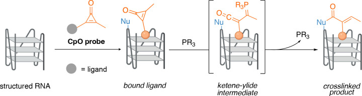
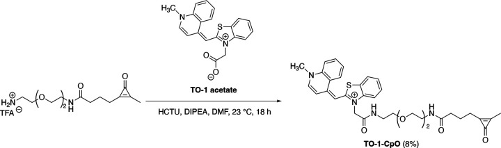
RNA sequences encode secondary and tertiary structures that impact protein production and other cellular processes. Misfolded RNAs can also potentiate disease, but the complete picture is lacking. To establish more comprehensive and accurate RNA structure-function relationships, new methods are needed to interrogate RNA and trap native conformations in cellular environments. Existing tools primarily rely on electrophiles that are constitutively "on" or triggered by UV light, often resulting in high background reactivity. We developed an alternative, chemically triggered approach to crosslink RNAs using bioorthogonal cyclopropenones (CpOs). These reagents selectively react with phosphines to provide ketenes-electrophiles that can trap neighboring nucleophiles to forge covalent crosslinks. As proof-of-concept, we synthesized a panel of CpOs and appended them to thiazole orange (TO-1). The TO-1 conjugates bound selectively to a model RNA aptamer (Mango) with nanomolar affinity, confirmed by fluorescence turn-on. After phosphine administration, covalent crosslinks were formed between the CpO probes and RNA. The degree of crosslinking was both time and dose-dependent. We further applied the chemically triggered tools to model RNAs in biologically relevant conditions. Collectively, this work expands the toolkit of probes for studying RNA and its native conformations.


Conflict of interest statement

The authors declare no competing financial interest.

Figures

Figure 1.
Figure 1.
Cyclopropenones as chemically triggered probes for examining RNA structure. In this work, the Mango aptamer was used as a model to demonstrate CpO-mediated crosslinking.
Figure 2.
Figure 2.. TO-1-CpO is activated upon phosphine administration and trapped by nucleophiles.
(A) Overall design of in vitro trapping experiments. (B-C) HPLC traces of TO-1-CpO and the quenched and crosslinked products. TO-1-CpO was incubated with PTA without any nucleophile to determine the retention time of hydrolyzed product. Ketene-ylide intermediates were trapped with (B) aniline or (C) benzylamine to forge covalent linkages.
Figure 3.
Figure 3.. TO-1-CpO binds Mango II with nanomolar affinity.
(A) Crystal structures of each Mango aptamer with TO-1-biotin depicted as space filling models. Mango I (PDB: 5V3F), Mango II (PDB: 6C63), Mango III (PDB: 6E8S), Mango IV (PDB: 6V9D). (B) Fluorescence turn-on analyses of Mango II samples incubated with TO-1-biotin, TO-1-CpO, or no reagent. (C) Fluorescence measurements resulting from TO-1-CpO (0–10 μM) incubated with each Mango aptamer. These data were used to calculate binding affinity (KD) of TO-1-CpO for each flavor of Mango, reported below the graph. Error bars represent the standard error of the mean for n = 3 experiments.
Figure 4.
Figure 4.. CpO-mediated crosslinking of Mango II is dependent on ligand concentration, time, and phosphine nucleophilicity.
(A) Overall scheme of TO-1-CpO crosslinking. The ligand was incubated with Mango aptamers for 10 min, prior to phosphine addition. (B) Denaturing PAGE analysis of crosslinking reactions. 5′-Cy5 labeled Mango II (10 μM) was incubated with varying concentrations of TO-1-CpO and PTA (10 mM). (C) Denaturing PAGE analysis of crosslinking time dependence. 5′-Cy5 labeled Mango II (10 μM) was incubated with TO-1-CpO (250 μM) and PTA (10 mM), and samples were analyzed over 5 h. (D) Quantification of relative crosslinking yields with various phosphine triggers. 5′-Cy5 labeled Mango II (10 μM) was incubated for 2 h with various phosphines (10 mM), and reaction products were analyzed via denaturing PAGE. Error bars represent the standard error of the mean for independent replicate experiments (n = 4 for PTA and n = 3 for TPTPS). (E) Denaturing PAGE analysis of crosslinking experiments performed in the presence of competing ligand (TO-1-biotin). TO-1-biotin (75–500 μM) was added to reactions comprising Mango II (10 μM), TO-1-CpO (150 μM) and PTA (10 mM).
Figure 5.
Figure 5.
Proximity-dependent crosslinking was observed with TO-1-CpO and Mango II. (A) Crystal structure of Mango II aptamer docked with truncated TO-1-biotin. Approximate position of the CpO motif is highlighted in red. Sites of reactivity are colored in blue. (B) Close up of the binding domain highlighting the five modified bases. (C) Sequencing analyses of reverse transcribed Mango II aptamer. RT stops observed with TO-1-CpO modified Mango II aptamer (blue) were significantly higher at five sites in comparison to the unmodified RNA (orange).
Scheme 1.
Scheme 1.
Synthesis of TO-1-CpO

Similar articles

References

    1. Alberts B.; Johnson A.; Lewis J.; Raff M.; Roberts K.; Walter P. From RNA to Protein. In Molecular Biology of the Cell. 4th edition; Garland Science, 2002.
    1. Meyer S. M.; Williams C. C.; Akahori Y.; Tanaka T.; Aikawa H.; Tong Y.; Childs-Disney J. L.; Disney M. D. Small Molecule Recognition of Disease-Relevant RNA Structures. Chem. Soc. Rev. 2020, 49 (19), 7167–7199. 10.1039/D0CS00560F. - DOI - PMC - PubMed
    1. Sharp P. A. The Centrality of RNA. Cell 2009, 136 (4), 577–580. 10.1016/j.cell.2009.02.007. - DOI - PubMed
    1. Ganser L. R.; Kelly M. L.; Herschlag D.; Al-Hashimi H. M. The Roles of Structural Dynamics in the Cellular Functions of RNAs. Nat. Rev. Mol. Cell Biol. 2019, 20 (8), 474–489. 10.1038/s41580-019-0136-0. - DOI - PMC - PubMed
    1. Mustoe A. M.; Brooks C. L.; Al-Hashimi H. M. Hierarchy of RNA Functional Dynamics. Annu. Rev. Biochem. 2014, 83 (Volume 83, 2014), 441–466. 10.1146/annurev-biochem-060713-035524. - DOI - PMC - PubMed

Publication types