[Preprint]. 2024 Dec 21:2024.10.18.619149.
doi: 10.1101/2024.10.18.619149.

Computational design of bifaceted protein nanomaterials

Affiliations

Computational design of bifaceted protein nanomaterials

Sanela Rankovic et al. bioRxiv. .

Update in

  • Computational design of bifaceted protein nanomaterials.
    Rankovic S, Carr KD, Decarreau J, Skotheim R, Kibler RD, Ols S, Lee S, Chun JH, Tooley MR, Dauparas J, Eisenach HE, Glögl M, Weidle C, Borst AJ, Baker D, King NP. Rankovic S, et al. Nat Mater. 2025 Jul 31:10.1038/s41563-025-02295-7. doi: 10.1038/s41563-025-02295-7. Online ahead of print. Nat Mater. 2025. PMID: 40745094 Free PMC article.

Abstract

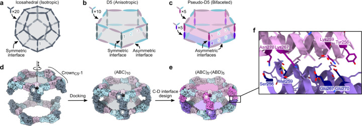
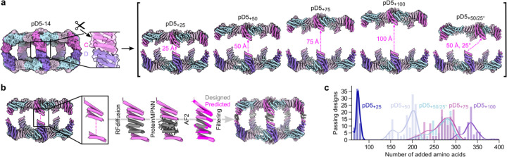
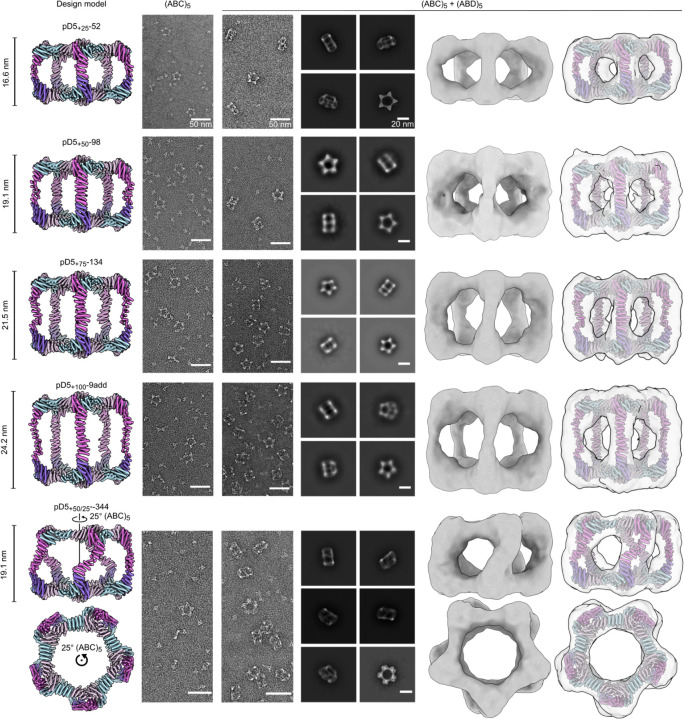
Recent advances in computational methods have led to considerable progress in the design of self-assembling protein nanoparticles. However, nearly all nanoparticles designed to date exhibit strict point group symmetry, with each subunit occupying an identical, symmetrically related environment. This limits the structural diversity that can be achieved and precludes anisotropic functionalization. Here, we describe a general computational strategy for designing multi-component bifaceted protein nanomaterials with two distinctly addressable sides. The method centers on docking pseudosymmetric heterooligomeric building blocks in architectures with dihedral symmetry and designing an asymmetric protein-protein interface between them. We used this approach to obtain an initial 30-subunit assembly with pseudo-D5 symmetry, and then generated an additional 15 variants in which we controllably altered the size and morphology of the bifaceted nanoparticles by designing de novo extensions to one of the subunits. Functionalization of the two distinct faces of the nanoparticles with de novo protein minibinders enabled specific colocalization of two populations of polystyrene microparticles coated with target protein receptors. The ability to accurately design anisotropic protein nanomaterials with precisely tunable structures and functions could be broadly useful in applications that require colocalizing two or more distinct target moieties.


Conflict of interest statement

Competing interests The authors declare no competing interests.

Figures

Figure 1.
Figure 1.. Overview of bifaceted pseudo-D5 architecture and design approach.
a, A nanoparticle with icosahedral symmetry constructed from homotrimeric building blocks requires only a single (symmetric) designed interface and is isotropic overall. b, A nanoparticle with dihedral symmetry (Dn; in this example n=5) constructed from a single heterotrimeric building block requires two designed interfaces: an asymmetric interface around the n-fold symmetry axis and a symmetric interface along the dihedral two-fold axes. The assembly is anisotropic, but the two opposing faces are constructed from the same three subunits (labeled 1, 2, and 3) and are not independently addressable. c, An anisotropic Dn assembly can be converted to a bifaceted pseudo-Dn assembly by asymmetrizing the designed interface at the dihedral two-fold axes. The nanoparticle is constructed from two different heterotrimers that can be produced independently prior to nanoparticle assembly, rendering each of the six subunits uniquely addressable, even if subunits 2+6 and 3+5 are genetically identical. d, Schematic of the procedure for docking CrownC5-1 substructures into (ABC)10 assemblies with D5 symmetry. The A, B, and C subunits of CrownC5-1 are labeled, and the rotational and translational degrees of freedom sampled during docking are indicated along the five-fold symmetry axis. e, An example bifaceted (ABC)5-(ABD)5 assembly after asymmetric C-D interface design. f, Details of an example asymmetric C-D interface. Positions featuring different amino acids in the C and D subunits are highlighted.
Figure 2.
Figure 2.. In vitro assembly and structural characterization of pD5–14.
a, SEC traces of pD5–14 (ABC)5, (ABD)5, and (ABC)5-(ABD)5 assemblies. b, SDS-PAGE of SEC-purified assemblies from panel (a). The locations of the A, B, C, and D subunits, as well as those of marker bands, are indicated. c–e, nsEM of pD5–14 (ABC)5 (c), (ABD)5 (d), and (ABC)5-(ABD)5 (e) assemblies, including representative 2D class averages and rigid-body fits of the computational design models into 3D reconstructions. f, Cryo-EM 2D class averages of pD5–14 (ABC)5-(ABD)5. g, Left: 4.30 Å resolution cryo-EM density map of pD5–14 (ABC)5-(ABD)5 viewed along two orthogonal axes. Right: Overlay between the computational design model (gray) and the experimentally determined cryo-EM model (colors). h, Details of asymmetrically designed C-D interface. Scale bars: 50 nm (raw micrographs) and 20 nm (2D class averages).
Figure 3.
Figure 3.. Fine-tuning of bifaceted protein nanoparticle size and shape using RFdiffusion.
a, Schematic of the approach used to define target extended architectures. Left: The scissors indicate the loop in the C subunit that was “cut”. Right: Target architectures were defined by translating and in one case rotating most of the (ABC)5 substructure while leaving the designed asymmetric C-D interface intact. b, Schematic of the design process used to generate de novo C subunits for each extended architecture. c, Number of designs that passed AF2 filtering (pLDDT > 90; RMSD to design model < 1.5 Å) per number of added amino acid residues for each extended architecture.
Figure 4.
Figure 4.. nsEM characterization of extended (ABC)5-(ABD)5 nanoparticles.
From top to bottom: Representative assemblies extended by 25, 50, 75, and 100 Å, or extended by 50 Å and rotated by 25°. From left to right: computational design models, raw micrographs of (ABC)5, raw micrographs of (ABC)5+(ABD)5, 2D class averages of (ABC)5+(ABD)5 showing representative top and side views, 3D reconstructions, and design models fit into the 3D reconstructions. Side views of the design models and 3D reconstructions are shown for each particle except pD5+50/25°-344, for which side and top views are shown. Scale bars: 50 nm (raw micrographs) and 20 nm (2D class averages).
Figure 5.
Figure 5.. Minibinder-functionalized bifaceted nanoparticles colocalize two distinct fluorescent polystyrene microparticle populations.
a, Schematic of the approach used to i) reorient the N termini of the A, B, and C subunits of pD5–14 to face toward the nanoparticle exterior and ii) independently functionalize each face of the resultant bifaceted nanoparticle with protein minibinders. The newly diffused ɑ-helix is shown in dark gray, and N and C termini are indicated by blue and red circles, respectively. b,c, SEC trace (b) and nsEM (c) of pD5–14_rd47 with Neo-2/15 and 41bb_mb1 genetically fused to the B components of the (ABC)5 and (ABD)5 components, respectively. Scale bars: 50 nm (raw micrograph) and 20 nm (2D class averages). d, Binding of functionalized bifaceted nanoparticles to IL-2Rβ (left) and 4–1BB (right) ectodomains, measured by BLI. The legend indicates which minibinder is fused to each component in each bifaceted nanoparticle. e, Representative flow cytometry plots showing colocalization of IL-2Rβ–coated Nile Red and 4–1BB–coated Purple Polystyrene particles by functionalized bifaceted nanoparticles. f, Quantitation of colocalization detected by flow cytometry. Each point represents an independent measurement, and the height of each bar indicates the mean for each group. Statistical significance was calculated via one-way ANOVA with Geisser-Greenhouse correction followed by Tukey’s multiple comparisons test, with individual variances computed for each comparison. *, p < 0.1; ****, p < 0.0001. g, Representative fluorescence microscopy images showing colocalization of IL-2Rβ–coated Nile Red and 4–1BB–coated Purple Polystyrene particles by functionalized bifaceted nanoparticles. The percentage of colocalized beads observed across 25 independent fields of view is displayed in the upper right corner of each image. Scale bar: 60 μm.

Publication types