. 2024 Dec;11(47):e2406027.
doi: 10.1002/advs.202406027. Epub 2024 Nov 1.

A Core-Brush Nanoplatform with Enhanced Lubrication and Anti-Inflammatory Properties for Osteoarthritis Treatment

Affiliations

A Core-Brush Nanoplatform with Enhanced Lubrication and Anti-Inflammatory Properties for Osteoarthritis Treatment

Yingying Liu et al. Adv Sci (Weinh). 2024 Dec.

Abstract

Osteoarthritis (OA) is recognized as a highly friction-related joint disease primarily associated with increased joint friction and inflammation due to pro-inflammatory M1-type macrophage infiltration in the articular cavity. Therefore, strategies to simultaneously increase lubrication and relieve inflammation to remodel the damaged articular microenvironment are of great significance for enhancing its treatment. Herein, a multifunctional core-brush nanoplatform composed of a ROS-scavenging polydopamine-coated SiO2 core and lubrication-enhancing zwitterionic poly(2-methacryloyloxyethyl phosphorylcholine) (PMPC) brush and loaded with the anti-inflammatory drug curcumin by a reactive oxygen species (ROS)-liable conjugation (named as SiO2@PP-Cur) is rationally designed. Benefiting from the grafted zwitterionic PMPC brush, a tenacious hydration layer with enhanced lubricity for reducing joint abrasions is developed. More importantly, based on the mono-iodoacetic acid-induced arthritis (MIA) rat model, intra-articular injection of SiO2@PP-Cur nanoplatform can effectively alleviate articular inflammation via promoting macrophage polarization from the pro-inflammatory M1 to anti-inflammatory M2 state by activating the nuclear factor erythroid 2-related factor 2 (Nrf2) signaling pathway and attenuating the degradation of cartilage matrix, resulting in the remodeling of the damaged microenvironment into a pro-regenerative microenvironment. As a result, SiO2@PP-Cur can considerably inhibit OA progression. Therefore, the work may provide a novel strategy for the development of an advanced core-brush nanoplatform for enhanced OA therapy.

Keywords: controlled release; drug delivery; enhanced lubrication; inflammatory regulation; osteoarthritis.


Conflict of interest statement

The authors declare no conflict of interest.

Figures

Scheme 1
Scheme 1
The schematic diagram for the design and mechanistic routes of SiO2@PP‐Cur NPs for the treatment of OA.
Figure 1
Figure 1
Characterization of SiO2@PP‐Cur NPs. A,B) The FT‐IR and XPS spectra of SiO2@PDA, SiO2@PDA‐Br, SiO2@PP, and SiO2@PP‐Cur, respectively. C) Hydrodynamic diameters and the zeta‐potential of SiO2 and SiO2@PP‐Cur. D) Representative TEM images of SiO2, SiO2@PDA, and SiO2@PP‐Cur. E) EDS elemental mapping for SiO2@PP‐Cur.
Figure 2
Figure 2
In vitro assays of lubricity, drug release, and ROS scavenging properties. A) Friction coefficient‐time plots of HA, SiO2, and SiO2@PP‐Cur. B) Lubrication properties of different concentrations of SiO2@PP‐Cur. C) Schematic illustration of the lubricating performance of SiO2@PP‐Cur. D) Release profiles of SiO2@PP‐Cur NPs in PBS in the presence or absence of 100 µm H2O2 at 37 °C. G) The influence of different nano‐formulations on the scavenging effect of H2O2 under identical conditions. UV–vis absorbance spectra of E) DPPH·, F) ABTS+·, H) O2 ·−, I) ·OH radicals after incubation with different nano‐formulations under identical conditions.
Figure 3
Figure 3
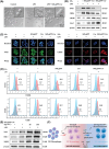
Assays on the anti‐inflammatory and anti‐oxidative efficacy of SiO2@PP‐Cur NPs. A) Protein expression levels of COX‐2 based on Western blot analysis of RAW264.7 cells supplemented with NPs for 24 h (SiO2@PP‐Cur: 50 µg·mL−1, n = 3). B) Confocal laser scanning microscope (CLSM) images of macrophages receiving different treatments for 24 h (green: DCFH; blue: nuclei) (n = 3). Flow cytometry analysis of C) ROS levels in macrophages 24 h post‐incubation with various reagents (n = 3), and D) the number of apoptotic cells in macrophages incubated for 24 h with various reagents (n = 3).
Figure 4
Figure 4
Macrophage phenotype reprogramming. A) Optical microscopic images comparing morphological characteristics of macrophages and LPS‐induced macrophages with or without SiO2@PP‐Cur treatment for 48 h. B) Relative protein expression of M1‐related markers and M2‐specific markers in M1‐polarized macrophages following incubation with different groups for 24 h (n = 3). C) Immunofluorescence staining images of LPS‐induced macrophages incubated for 24 h with various treatments (red: CD206; green: iNOS; blue: nuclei) (n = 3). D) Representative flow cytometry results of CD86 and CD206 expression levels in macrophages incubated for 24 h with various reagents (n = 3). E) Protein expression levels of Nrf2, HO‐1, and NQO‐1 in macrophages as identified by western blotting (n = 3). F) The possible anti‐inflammatory mechanism and macrophage phenotypic reprogramming.
Figure 5
Figure 5
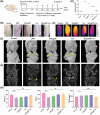
Morphological analysis of knee joints of OA rats after treatment with SiO2@PP‐Cur. A) Schematic diagram of the protocol for the construction and treatment of rat OA models. B) Representative digital images and C) thermographic images of joint knees at week 7. D) Quantification of the temperature of the knee joint in the various groups (n = 3). E,F) Representative CT scans of healthy and arthritic knees at week 7 (n = 3). G) BV/TV quantitative analysis, Tb.Th and Th.Sp of knee joints. Data are expressed as means ± SD. (n = 3), p* < 0.05, p**< 0.01, p***< 0.005.
Figure 6
Figure 6
Histological analysis of OA knee joints of rats following supplementation with SiO2@PP‐Cur. A) Representative H&E staining and B) Safranin O‐fast green staining of the cartilage sections of osteoarthritic rats following different treatments. C) Representative sections of immunohistochemical staining for collagen II. D) Representative sections of TUNEL staining for apoptotic cells. E) Heatmap of variables of histological scores. F) Hot plate test of rats at 7 weeks. G) OARSI grades of rat joints at 7 weeks. Data are presented as means ± SD. (n = 3), p* < 0.05, p**< 0.01, p***< 0.005.
Figure 7
Figure 7
A) H&E staining of synovial tissue. Synovial macrophages stained with B) iNOS and C) CD206 markers. D) Serum levels of TNF‐α, IL‐6, and IL‐1β in the different groups. Data are presented as means ± SD. (n = 3), p* < 0.05, p**< 0.01, p***< 0.005.

Similar articles

References

    1. Wen J., Li H., Dai H., Hua S., Long X., Li H., Ivanovski S., Xu C., Mater. Today Bio. 2023, 19, 100597. - PMC - PubMed
    1. Zhang Y., Liu Y., Hou M., Xia X., Liu J., Xu Y., Shi Q., Zhang Z., Wang L., Shen Y., Yang H., He F., Zhu X., Adv. Sci. 2023, 10, 2206144. - PMC - PubMed
    1. Han Z., Bai L., Zhou J., Qian Y., Tang Y., Han Q., Zhang X., Zhang M., Yang X., Cui W., Hao Y., Biomaterials. 2022, 285, 121545. - PubMed
    1. Bruno M. C., Cristiano M. C., Celia C., d'Avanzo N., Mancuso A., Paolino D., Wolfram J., Fresta M., ACS Nano. 2022, 16, 19665. - PubMed
    1. Qiu W., Zhao W., Zhang L., Wang H., Li N., Chen K., Zhang H., Wang Y., Adv. Funct. Mater. 2022, 32, 2208189.

LinkOut - more resources